Classical X Ray Signs in congenital heart disease.

By Dr Deepu
X ray finding: Boot shaped heart
Condition: TOF(tetrology of Fallot)


X ray finding:Goose neck sign
Condition:Endocardial Cushion defect


X ray finding:Figure of 3/reverse figure of 3
Condition: Coarctation of Aorta


X ray finding: Box shaped heart
Condition :Ebsteins anomaly


X ray finding: Egg on String/egg on side
Condition: Transposition of great vessels


X ray finding: Figure of 8/ snowman
Condition: total anomalous Pulmonary venous circulation


X ray finding: Scimitar sign
Condition: partial anomalous pulmonary venous return


Image credit: Facebook group pgblaster India
https://m.facebook.com/PgblasterIndia/

Pneumothorax Signs

By Dr Deepu
ANS--1.pneumothorax


Atypical signs of pneumothorax


Atypical sign  arise when the patient is supine or the pleural space partly obliterated.
In the supine position, pleural air rises and collects anteriorly, particularly medially and basally, and may not extend far enough posteriorly to separate lung from thechest wall at the apex or laterally.


Signs that suggest a pneumothorax under these conditions are

1.ipsilateral transradiancy, either generalized or hypochondrial
 2.a deep, finger-like costophrenic sulcus laterally
3. a visible anterior costophrenic recess seen as an oblique line or interface in the hypochondrium; when the recess is manifest as an interface it mimics the adjacent diaphragm (‘double diaphragm sign’)
4.a transradiant band parallel to the diaphragm and/or mediastinum with undue clarity of the mediastinal border
5. visualization of the undersurface of the heart, and of the cardiac fat pads as rounded opacities suggesting masses
6.diaphragm  depression.

MCQ Double Diaphragm Sign

By Dr Deepu
Double diaphragm sign

Q1.' Double diaphragm sign 'is noted in
a.subpulmonic effusion
b.pneumothorax
c.hydrothorax
d.diaphragmatic palsy

Answer click here

Lung Collapse

By Dr Deepu
LEFT UPPER LOBE COLLAPSE


RADIOGRAPHIC CONSIDERATIONS FOR COLLAPSE OF LUNG


The cardinal radiographic features of lobar collapse are
1. increased opacity of the affected lobe-A collapsed lobe appears radiographically dense due to a combination of retained secretions or fluid within the lobe and reduction in aeration of the lobe
2.volume loss - can be inferred by direct and indirect signs.
A.Direct signs of volume loss refer to displacement of interlobar fissures, crowding of  pulmonary vessels and bronchi.  Hilar elevation on the PA chest radiograph is a well-known sign of upper lobe collapse:
B. indirect signs include compensatory shifts of adjacent structures such as hyperinflation of other lobes and mediastinal shift (trachea etc)
  The normal lung parenchyma should expand proportionally to compensate for the degree of collapse and often the greater the degree of lobar collapse, the greater the compensatory overinflation.
Therefore when small lung volumes are involved, the hyperinflation usually only involves the remainder of the ipsilateral lung, whereas with larger volumes, the contralateral lung may expand across the midline.
Shifting granuloma sign ----Hyperexpansion may also result in a change in position of lung lesions, such as granulomas resulting in the so-called shifting granuloma sign
The Luftsichel sign (from German, meaning air crescent)is a particular manifestation of the hyperexpansion. It is due to the overinflated superior segment of the ipsilateral lower lobe occupying the space between the mediastinum and the medial aspect of the collapsed upper lobe, resulting in a paramediastinal translucency . The sign is more common on the left than the right and is regarded as a typical appearance of left upper lobe collapse

Juxtaphrenic peak of the diaphragm ---- A useful ancillary sign of upper lobe collapse (or a combination of right upper and middle lobe collapse) is a juxtaphrenic peak of the diaphragm . The sign refers to a small triangular density at the highest point of the dome of the hemidiaphragm, due to the anterior volume loss of the affected upper lobe resulting in traction and reorientation of an inferior accessory fissure

Golden's S sign ---The sign refers to the S shape (or more accurately, reverse S on the right) of the fissure due to the combination of collapse and mass centrally resulting in a focal convexity with a concave outline peripherally.Lobar collapse due to a central obstructing bronchogenic carcinoma is most likely when Golden's S sign is seen.

MCQ - Luftsichel sign.

By Dr Deepu

Q.Paramediastinal translucency(Luftischel’s sign) is typically seen in
a.left upper lobe collapse
b.left lower lobe collapse
c.right lower lobe collapse
d.right middle lobe collapse

Answer click here

Chest challenge answer

By Dr Deepu
The Answer is D.
Explanation
The scenario is of decreased lung complaince that is restrictive lung disease.
In restrictive lung disease, it may be intrathoracic (pulmonary fibrosis) or extrathoracic ( like ALS, myaesthenia gravis). It can be differentiated by DLCO which will be decreased in fibrosis and
normal in extrathoracic causes.

Chest challenge -Difficulty ventilating

By Dr Deepu

A 55-year-old male with chronic dyspnea is intubated and started on mechanical ventilation for respiratory failure Pressure-volume scalars on the ventilator show decreased change in volume for each unit change in pressure as compared to normal values. Of the following options, which is the most likely cause of  his respiratory failure?

A. Amyotrophic lateral sclerosis

B.Asthma

C. Centriacinar emphysema

D. Pulmonary fibrosis

E. Alpha-1 -antitrypsin deficiency

Click for answer

FDA approves cancer drugs with no evidence of longer life or better quality of life, study says

By Dr Deepu

 A new study published in the Mayo Clinic Proceedings “says the FDA has been approving cancer drugs that may have no scientific correlation to actually living longer or maintaining quality of life.” According to the study, the FDA’s use of surrogate measures often do not reflect an increase in overall survival or quality of life. The study found that “among 25 drugs approved under the accelerated approval program between 2009 and 2014, 14 – or 56% – did not have supporting evidence that they lengthened life or maintained quality of life,” while 37% of drugs approved under traditional approval “were similarly lacking.”

The Luftsichel Sign

By Dr Deepu


 The Luftsichel sign is seen in some cases of left upper lobe collapse and refers to the frontal chest radiographic appearance due to hyperinflation of the superior segment of the left lower lobe interposing itself between the mediastinum and the collapsed left upper lobe.

Radiographic features

Chest radiograph

In many cases of left upper lobe collapse the anterior parts of the aortic arch, and thus the aortic knuckle, are abutted by collapsed lung and thus the normal silhouette is lost. In some case the apical (superior) segment of the left lower lobe is hyperinflated and becomes interposed between the collapsed lung and the adjacent aortic arch. In such cases the aortic knuckle silhouette remains visible. The collapsed left upper lobe is thus displaced laterally away from the mediastinum.
Luftsichel sign ( A german word ; Luft = Air and Sichel = Sickle ) . The luftsichel sign represents the hyperexpanded superior segment of the left lower lobe interposed between the atelectatic left upper lobe and the aortic arch.

Reference:

Webb WR, Higgins CB. Thoracic Imaging: Pulmonary and Cardiovascular Radiology. Lippincott, Williams & Wilkins 2005
http://radiopaedia.org/articles/luftsichel-sign
http://www.radpod.org/2007/06/08/left-upper-lobe-collapse/
http://www.lungindia.com/article.asp?issn=0970-2113;year=2012;volume=29;issue=1;spage=83;epage=84;aulast=Singh
http://www.radrounds.com/photo/luftsichel-sign

Bayer withdraws Phase 2 Trial of PH drug in IPF due to death risk

By Dr Deepu


Potentially higher risk of death seen in people with rare disease that has no approved treatments

Bayer has stopped a Phase 2 clinical trial (NCT02138825) evaluating riociguat (Adempas) in patients with pulmonary hypertension associated with idiopathic interstitial pneumonias (PH-IIP) on the recommendation of the study’s Data Monitoring Committee (DMC).
Download the ChestMedicine.org free app from playstore


The committee — sometimes called a data and safety monitoring board — is an independent group of experts who monitor patient safety and treatment efficacy data during a clinical trial. While reviewing the riociguat data, the DMC determined that patients receiving this treatment were at a potentially higher risk of death and other serious adverse events compared to those receiving a placebo. The DMC did not find any particular cause or common characteristic in the patients who died, but many were found to have more advanced lung disease than the clinical trial cohort as a whole.

People taking part in the trial will be monitored for safety for a minimum of four months after discontinuing treatment, Bayer said.

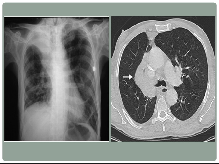
PH-IIP is a severe and rare disease. Pulmonary hypertension (PH) is an increase of blood pressure in the pulmonary artery, pulmonary vein, or pulmonary capillaries, together known as the lung vasculature, leading to shortness of breath, dizziness, leg swelling, and other symptoms. PH is increasingly recognized as a complication of interstitial lung disease (ILD). It can exist when ILD is mild, but is more common when hypoxemia and severe pulmonary dysfunction exist.

IIPs are ILDs of unknown etiology that share similar clinical and radiologic features, and are distinguished primarily by the histopathologic patterns found in a lung biopsy, being characterized by varying degrees of inflammation and fibrosis. All IIPs cause dyspnea. Treatment varies by subtype, as does disease prognosis.

Patients with PH-IIP are a high-risk patient population with an estimated mortality rate of over 20% within one year. Currently, there are no approved treatments for PH-IIP.

Riociguat’s positive benefit-risk profile for its approved indications has not been altered, including its use to treat pulmonary hypertension patients in WHO Groups 1 and 4. Bayer announced that it is carefully examining riociguat’s efficacy and safety on an ongoing basis.

“We understand that the need to terminate the study in PH-IIP is very disappointing for patients suffering from this disease, as well as for their doctors and healthcare providers. There is a significant unmet medical need for PH-IIP patients as there are no approved treatments, and finding an effective treatment remains a challenge,” said Dr. Joerg Moeller, a member of the Bayer Pharmaceuticals Executive Committee and head of Global Development, in a press release. “Bayer remains committed to identifying new therapeutic options and to improving the lives of patients in disease areas where there is a high unmet medical need such as pulmonary hypertension.”

Adempas is Bayer’s novel formulation to treat PH patients. Chemically known as riociguat, the drug is the first of a new class of stimulators of soluble guanylate cyclase (sGC), an enzyme responsible for vasodilation and lowering of blood pressure, and the only receptor of nitric oxide in the body.

3D printing technology may change the way we treat patients

By Dr Deepu

3D printing technology has been in existence since the 1980s. Charles “Chuck” Hull, cofounder of 3D Systems, is credited with the invention of the world’s first 3D printer (stereolithography) in 1983. In the mid to late 1980s, there was a proliferation of 3D printing technology. In 1987, Dr Carl Deckard developed the selective laser sintering (SLS) process. In 1989, Scott Crump invented fusion deposition modeling and went on to cofound Stratasys. Today, these two companies, 3D Systems and Stratasys, are the leaders in the 3D printing industry.

With three-dimensional (3D) printers, digital surface models are readily made into physical models to allow rapid prototyping. 3D printing has been increasingly applied to medical disciplines in which therapeutic interventions heavily depend on appreciation of complex anatomic structural relationships.
The anatomy of the tracheobronchial tree is uniquely suited for 3D printing technology. The adult trachea has an internal diameter of approximately 16 to 20 mm and spans approximately 10 to 13 cm. It is covered with 16 to 20 C-shaped pieces of cartilage anterolaterally, and the posterior trachea is membranous. The right mainstem bronchus is approximately 1.5 cm long with an internal diameter of 10 to 12 mm. The left mainstem bronchus is approximately 4 to 4.5 cm long with an internal diameter of 8 to 12 mm (DICOM dataset file available online as supplementary material). The current molded silicone stents (DUMON; Novatech) have the appropriate ranges of diameter (9-18 mm), thickness (1-1.5 mm), and length (20-110 mm). These stents are manufactured with a conventional mold injection technique and come in predefined sizes. If custom modifications were needed, a custom stent could be generated in approximately 3 weeks, a substantial wait time for the patient.

HIlum Overlay Sign

By Dr Arun M, KMC Mangalore

Halo Sign

By Dr Arun M, KMC Mangalore


Hamptons Hump

By Dr Arun M, KMC Mangalore


Golden S Sign

By Dr Arun M, KMC Mangalore


Gloved finger sign

By Dr Arun M, KMC Mangalore

Galaxy Sign

By Dr Arun M, KMC Mangalore


Fleischners Sign

By Dr Arun M, KMC Mangalore


Flat Waist Sign

By Dr Arun M, KMC Mangalore

Feeding vessel Sign

By Dr Arun M, KMC Mangalore