Chest Medicine Challenge-02, Case of The week ( 11-17th April)- Answer

Pulmonary Medicine blog

Its Tuesday and here is the answer for the chest Medicine challenge 2.
Raise your hand if you answered D to Friday's CHEST Challenge question! Nice work! Read on for complete rationale.
A 45-year-old nonsmoking woman was referred for an opinion regarding management of recurrent pneumothorax. She was well until age 27 when she had a right-sided spontaneous pneumothorax. Two years later, she had another right-sided pneumothorax and underwent thoracotomy and stapling of the right lung apex. She has had no further episodes since that time. She is seeing a dermatologist for multiple facial papules, but otherwise, her general health is excellent. She denies any respiratory symptoms. Results of a physical examination are normal other than multiple skin-colored papules over the 
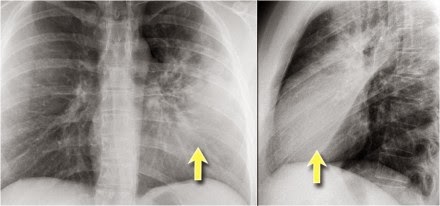
central face and nose. Results of a chest radiograph demonstrated right apical pleural thickening but were otherwise normal. A chest CT was performed and representative images are shown in Figures 138-A and 138-B. Her lung function was normal. The most likely diagnosis of this condition is:
A. Langerhans cell histiocytosis.
B. Lymphangioleiomyomatosis (LAM).
C. Sarcoidosis.
D. Birt-Hogg-Dubé syndrome (BHDS).


The CT scan reveals several cysts in the left lung and scarring at the right apex related to the previous surgery. A history of recurrent pneumothorax, lung cysts, and skin lesions (fibrofolliculomas) with normal lung function is consistent with the diagnosis of Birt-Hogg-Dubé syndrome (BHDS) (choice D is correct). Langerhans cell histiocytosis is characterized by diffuse cystic disease of the lung, spontaneous pneumothorax, and airway obstruction related to cigarette smoking. These findings are not present in this patient (choice A is incorrect). Similarly, airflow obstruction, recurrent pleural effusions, and diff use pulmonary disease are characteristic of LAM, features that also are not found in this patient (choice B is incorrect). Sarcoidosis is a granulomatous inflammatory lung disease characterized by diff use parenchymal opacities, airflow obstruction, and possibly, skin lesions. Lupus pernio, one of the skin manifestations of sarcoidosis, appears as purple nodules on the nose, cheeks, and ears, and none of these features is present in this patient (choice C is incorrect).
BHDS is an autosomal dominantly inherited genodermatosis that predisposes a person to the development of cutaneous hamartomas (fibrofolliculomas), kidney neoplasms, lung cysts, and spontaneous pneumothorax. The BHD locus has been mapped to the short arm of chromosome 17(17p11.2). BHD is composed of 14 exons, and more than 40 unique mutations in BHD have been reported. Most BHD germline mutations are frameshift or nonsense mutations that are predicted to truncate the BHD protein, folliculin. Patients exhibit multiple 1- to 5-mm white-colored or skin-coloured papules distributed over the face, neck, and/or upper trunk, which histologically, are fibrofolliculomas (Fig 138-C). BHDS is associated with a unique histologic spectrum of bilateral and multifocal kidney tumors ranging from hybrid oncocytic (67%) to chromophobe renal carcinoma (23%) to oncocytic renal carcinoma (3%). Clear cell renal cell carcinoma (3%) has also been reported in a few patients with BHDS. Pulmonary manifestations are a major feature of BHDS. Most patients who are affected (89%) have multiple pulmonary cysts. The number of lung cysts is clearly related to the risk of pneumothorax. Approximately one-quarter of patients with BHDS have a history of one or more pneumothoraces. Smoking does not appear to be a risk factor for pneumothorax in this population.
Toro JR, Wei1 MH, Glenn GM, et al. BHD mutations, clinical and molecular genetic investigations of Birt-Hogg-Dubé syndrome: a new series of 50 families and a review of published reports. J Med Genet. 2008;45(6):321-331.
Toro JR, Pautler SE, Stewart L, et al. Lung cysts, spontaneous pneumothorax, and genetic associations in 89 families with Birt-Hogg-Dubé syndrome. Am J Respir Crit Care Med. 2007;175(10):1044-1053.

Pavlovich CP, Grubb RL III, Hurley K, et al. Evaluation and management of renal tumors in the Birt-Hogg-Dubé syndrome. J Urol. 2005; 173:1482-1486.

RESPIRATORY FAILURE

Pulmonary Medicine blog

Respiratory failure occurs when the respiratory system fails in oxygenation and/or carbon dioxide (CO2) elimination. Respiratory failure may be:[1]
  • Hypoxaemic respiratory failure (type I respiratory failure): PaO2 is less than 60 mm Hg (8 kPa) with a normal or low PaCO2. This is caused by ventilation-perfusion mismatch with either/both:
    • Under-ventilated alveoli (eg pulmonary oedema, pneumonia or acute asthma).
    • Venous blood bypasses ventilated alveoli (eg right to left cardiac shunts).
    Hyperventilation increases CO2 removal but does not increase oxygenation, as blood leaving unaffected alveoli is almost fully saturated.
  • Hypercapnic respiratory failure (type II respiratory failure): PaCO2 is more than 50 mm Hg (6.5 kPa) and indicates inadequate alveolar ventilation. Any ventilation-perfusion mismatch will affect PaO2 and therefore hypoxaemia is also common.
Respiratory failure may be acute or chronic:[2]
  • Acute hypercapnic respiratory failure develops over minutes to hours. The pH is usually therefore less than 7.3.
  • Chronic respiratory failure develops over several days or longer. There is sufficient time for renal compensation and an increase in bicarbonate so the pH is usually only slightly decreased. Clinical markers of long-standing hypoxaemia include polycythaemia and cor pulmonale.

Common causes of type I respiratory failure

  • Chronic obstructive pulmonary disease (COPD).
  • Pneumonia.
  • Pulmonary oedema.
  • Pulmonary fibrosis.
  • Asthma.
  • Pneumothorax.
  • Pulmonary embolism.
  • Pulmonary hypertension.
  • Cyanotic congenital heart disease.
  • Bronchiectasis.
  • Acute respiratory distress syndrome.
  • Kyphoscoliosis.
  • Obesity

Common causes of type II respiratory failure

  • COPD.
  • Severe asthma.
  • Drug overdose, poisoning.
  • Myasthenia gravis.
  • Polyneuropathy.
  • Poliomyelitis.
  • Muscle disorders.
  • Head injuries and neck injuries.
  • Obesity.
  • Pulmonary oedema.
  • Adult respiratory distress syndrome.
  • Hypothyroidism.
The cause of respiratory failure is often clear from a thorough history and physical examination. See also the separate article Respiratory System - History and Examination.

Symptoms

  • The history may indicate the underlying cause, eg paroxysmal nocturnal dyspnoea, and orthopnoea in pulmonary oedema.
  • Both confusion and reduced consciousness may occur.

Signs

  • Localised pulmonary findings are determined by the underlying cause.
  • Neurological features may include restlessness, anxiety, confusion, seizures, or coma.
  • Tachycardia and cardiac arrhythmias may result from hypoxaemia and acidosis.
  • Cyanosis.
  • Polycythaemia is a complication of long-standing hypoxaemia.
  • Cor pulmonale: pulmonary hypertension is frequently present and may induce right ventricular failure, leading to hepatomegaly and peripheral oedema.
  • Arterial blood gas analysis: confirmation of the diagnosis.
  • CXR: often identifies the cause of respiratory failure.
  • FBC: anaemia can contribute to tissue hypoxia; polycythaemia may indicate chronic hypoxaemic respiratory failure.
  • Renal function tests and LFTs: may provide clues to the aetiology or identify complications associated with respiratory failure. Abnormalities in electrolytes such as potassium, magnesium, and phosphate may aggravate respiratory failure and other organ dysfunction.
  • Serum creatine kinase and troponin I: to help exclude recent myocardial infarction. Elevated creatine kinase may also indicate myositis.
  • TFTs (hypothyroidism may cause chronic hypercapnic respiratory failure).
  • Spirometry: useful in the evaluation of chronic respiratory failure.
  • Echocardiography: if a cardiac cause of acute respiratory failure is suspected.
  • Pulmonary function tests are useful in the evaluation of chronic respiratory failure.
  • ECG: to evaluate a cardiovascular cause; it may also detect dysrhythmias resulting from severe hypoxaemia or acidosis.
  • Right heart catheterisation: should be considered if there is uncertainty about cardiac function, adequacy of volume replacement, and systemic oxygen delivery.
  • Pulmonary capillary wedge pressure may be helpful in distinguishing cardiogenic from noncardiogenic oedema.
A patient with acute respiratory failure generally needs prompt hospital admission in an intensive care unit. Many patients with chronic respiratory failure can be treated at home, depending on the severity of respiratory failure, underlying cause, comorbidities and social circumstances.
  • Immediate resuscitation may be required.
  • Appropriate management of the underlying cause.

Hypoxaemia

  • Ensure adequate oxygen delivery to tissues, generally achieved with a PaO2 of 60 mm Hg or an arterial oxygen saturation (SaO2) of greater than 90%.
  • Beware the prolonged use of high-concentration oxygen in chronic sufferers who have become reliant on their hypoxic drive to maintain an adequate ventilation rate. Elevating the PaO2 too much may reduce the respiratory rate so that the PaCO2 may rise to dangerously high levels.
  • Assisted ventilation:
    • Mechanical ventilation is used to increase PaO2 and to lower PaCO2. Mechanical ventilation also rests the respiratory muscles and is an appropriate therapy for respiratory muscle fatigue. Weaning patients with chronic respiratory failure off of mechanical ventilation may be very difficult.[2]
    • Non-invasive ventilation (NIV) has been increasingly used as an alternative to intubation. NIV improves survival and reduces complications for selected patients with acute respiratory failure. The main indications are exacerbation of COPD, cardiogenic pulmonary oedema, pulmonary infiltrates in immunocompromised patients, and weaning of previously intubated stable patients with COPD.[3]

Hypercapnia and respiratory acidosis

Correct the underlying cause and/or provide assisted ventilation.
  • Pulmonary: for example, pulmonary embolism, pulmonary fibrosis, and complications secondary to the use of mechanical ventilation.
  • Cardiovascular: for example, cor pulmonale, hypotension, reduced cardiac output, arrhythmias, pericarditis, and acute myocardial infarction.
  • Gastrointestinal: for example, haemorrhage, gastric distention, ileus, diarrhoea, and pneumoperitoneum. Duodenal ulceration caused by stress is common in patients with acute respiratory failure.
  • Polycythaemia.
  • Hospital-acquired infection: for example, pneumonia, urinary tract infections, and catheter-related sepsis, are frequent complications of acute respiratory failure.
  • Renal: acute kidney injury (acute renal failure) and abnormalities of electrolytes andacid-base balance are common in critically ill patients with respiratory failure.
  • Nutritional: including malnutrition and complications related to administration of enteral or parenteral nutrition. Complications associated with nasogastric tubes, eg abdominal distention and diarrhoea.
The mortality rate associated with respiratory failure depends on the underlying cause.

Further reading & references

  1. Roussos C, Koutsoukou A; Respiratory failure. Eur Respir J Suppl. 2003 Nov;47:3s-14s.
  2. Kaynar AM et al, Respiratory Failure, Medscape, Sep 2010
  3. Nava S, Hill N; Non-invasive ventilation in acute respiratory failure. Lancet. 2009 Jul 18;374(9685):250-9.

BASICS OF CHEST X RAY-PART 4, THE HEART AND PERICARDIUM


This is the fourth Post in the series Chest X Ray Interpretation. I personally suggest to read the other three posts before proceeding

1

    Heart and Pericardium
    On a chest film only the outer contours of the heart are seen.
    In many cases we can only tell whether the heart figure is normal or enlarged and it will be difficult to say anything about the different heart compartments.

    IMPROVE YOUR X RAY READING SKILLS, BUY A BOOK


    However it can be helpful to know where the different compartments are situated.
    Left Atrium
        Most posterior structure.
        Receives blood from the pulmonary veins that run almost horizontally towards the left atrium.
        Left atrial appendage (in purple) can sometimes be seen as a small outpouching just below the pulmonary trunk.
        Enlargement of the left atrium results on the PA-view in outpouching of the upper heart contour on the right and an obtuse angle between the right and left main bronchus. On the lateral view bulging of the upper posterior contour will be seen.

    Right Atrium
        Receives blood from the inferior and superior vena cava.
        Enlargement will cause an outpouching of the right heart contour.

    Left Ventricle
        Situated to the left and posteriorly to the right ventricle.
        Enlargement will result on the PA-view in an increase of the heart size to the left and on the lateral view in bulging of the lower posterior contour.

    Right Ventricle
        Most anterior structure and is situated behind the sternum.
        Enlargement will result on the PA-view in an increase of the heart size to the left and can finally result in the left heart border being formed by the right ventricle.
    Left Atrium
        The upper posterior border of the heart is formed by the left atrium.
        Enlargement will result in bulging of the upper posterior contour

    Left Ventricle
        Forms the lower posterior border.
        Enlargement will displace the contour more posteriorly.

    Right Ventricle
        The lower retrosternal space is filled by the right ventricle.
        Enlargement of the right ventricle will result in more superior filling of this retrosternal space.

    Left Atrium enlargement
    This is a patient with longstanding mitral valve disease and mitral valve replacement.
    Extreme dilatation of the left atrium has resulted in bulging of the contours (blue and black arrows).

    Right ventricle enlargement
    First study the PA and lateral chest film and then continue reading.

    On these chest films the heart is extremely dilated.
    Notice that it is especially the right ventricle that is dilated. This is well seen on the lateral film (yellow arrow).

    There is a small aortic knob (blue arrow), while the pulmonary trunk and the right lower pulmonary artery are dilated.
    All these findings are probably the result of a left-to-right shunt with subsequent development of pulmonary hypertension.
    The location of the cardiac valves
    is best determined on the lateral radiograph.
    A line is drawn on the lateral radiograph from the carina to the cardiac apex.
    The pulmonic and aortic valves generally sit above this line and the tricuspid and mitral valves sit below this line .

    On this lateral view you can get a good impression of the enlargement of the left atrium.

    Cardiac incisura


    On the right side of the chest the lung will lie against the anterior chest wall.
    On the left however the inferior part of the lung may not reach the anterior chest wall, since the heart or pericardial fat or effusion is situated there.
    This causes a density on the anteroinferior side on the lateral view which can have many forms.
    It is a normal finding, which can be seen on many chest x-rays and should not be mistaken for pathology in the lingula or middle lobe.

    The explanation for the cardiac incisura is seen on this CT-image.
    At the level of the inferior part of the heart we can appreciate that the lower lobe of the right lung is seen more anteriorly compared to the left lower lobe.

    Pacemaker
    There are different types of cardiac pacemakers.
    Here we see a pacemaker with one lead in the right atrium and another in the right ventricle.

    A third lead is seen, which is guided through the coronary sinus towards the left ventricle.
    This is done in patients with asynchrone ventricular contractions.
    Pacing both ventricles at the same time will lead to synchrone contractions and a better cardiac output.


    Pericardial effusion
    Whenever we encounter a large heart figure, we should always be aware of the possibility of pericardial effusion simulating a large heart.

    On the chest x-ray it looks as if this patient has a dilated heart while on the CT it is clear, that it is the pericardial effusion that is responsible for the enlarged heart figure.
    Especially in patients who had recent cardiac surgery an enlargement of the heart figure can indicate pericardial bleeding.

    This patient had a change in the heart configuration and pericardial bleeding was suspected.
    Ultrasound demonstrated only a minimal pericardial effusion.
    Continue with the CT.

    There is a large pericardial effusion, which is located posteriorly to the left ventricle (blue arrow).
    The left ventricle id filled with contrast and is compressed (red arrow).
    At surgery a large hematoma in the posterior part of the pericardium was found.

    Notice that on the anterior side there is only a minimal collection of pericardial fluid, which explains why the ultrasound examination underestimated the amount of pericardial fluid.

    Here another patient who had valve-replacement.

    Notice the large heart size.
    There is redistribution of the pulmonary vessels which indicates heart failure.

    Continue with the CT.
      

    The CT-image shows a large pericardial effusion.

    Always compare these post-operative chest films with the pre-operative ones.

    Calcifications

    Detection of calcifications within the heart is quite common.
    The most common are coronary artery calcifications and valve calcifications.

    Here we see pericardial calcifications which can be associated with constrictive pericarditis.
    In this case there are calcifications that look like pericardial calcifications,
    but these are myocardial calcifications in an infarcted area of the left ventricle.

    Notice that they follow the contour of the left ventricle.

    Pericardial fatpad

    Pericardial fat depositions are common.
    Sometimes a large fat pad can be seen (figure).

    Necrosis of the fat pad has pathologic features similar to fat necrosis in epiploic appendagitis.
    It is an uncommon benign condition, that manifests as acute pleuritic chest pain in previously healthy persons.

    Pericardial cyst     


    Pericardial cysts are connected to the pericardium and usually contain clear fluid.
    The majority of pericardial cysts arise in the anterior cardiophrenic angle, more frequently on the rightside, but they can be seen as high as the pericardial recesses at the level of the proximal aorta and pulmonary arteries .
    Most patients are asymptomatic.
    On the chest x-ray it seems as if there is a elevated left hemidiaphragm.
    On CT however there is a cyst connected to the pericardium

    suggested reading


    WHO’s first global report on antibiotic resistance reveals serious, worldwide threat to public health

    New WHO report provides the most comprehensive picture of antibiotic resistance to date, with data from 114 countries

    News release  GENEVA -
    A new report by WHO–its first to look at antimicrobial resistance, including antibiotic resistance, globally–reveals that this serious threat is no longer a prediction for the future, it is happening right now in every region of the world and has the potential to affect anyone, of any age, in any country. Antibiotic resistance–when bacteria change so antibiotics no longer work in people who need them to treat infections–is now a major threat to public health. “Without urgent, coordinated action by many stakeholders, the world is headed for a post-antibiotic era, in which common infections and minor injuries which have been treatable for decades can once again kill,” says Dr Keiji Fukuda, WHO’s Assistant Director-General for Health Security. “Effective antibiotics have been one of the pillars allowing us to live longer, live healthier, and benefit from modern medicine. Unless we take significant actions to improve efforts to prevent infections and also change how we produce, prescribe and use antibiotics, the world will lose more and more of these global public health goods and the implications will be devastating.”
    read the complete article from WHO 

    Basics of Chest X Ray Part 3- systematic approach, silhouette sign and Hidden areas in chest Xray

    This is the third post in the series, this post deals with the systematic interpretation and analysis of X-ray Chest with special emphasis on silhouette sign and hidden areas of the lung.

    Whenever you review a chest x-ray, always use a systematic approach.
    We use an inside-out approach from central to peripheral.


    First the heart figure is evaluated, followed by mediastinum and hili.
    Subsequently the lungs, lungborders and finally the chest wall and abdomen are examined.

    You have to know the normal anatomy and variants.

    Find subtle abnormalities by using the sihouette sign and mediastinal lines.
    Once you see an abnormality use a pattern approach to come up with the most likely diagnosis and differential diagnosis.

    Old films
    It is extremely important to always compare with old films, as we will demonstrate in this case.
    Actually someone said that the most important radiograph is the old film, since it gives you so much information.
    For instance a lung mass, which hasn't changed in many years is not a lung cancer.
    First study the chest films.
    Then continue.
    Based on the CXR that you just saw, you could have made the diagnosis of congestive heart failure, but the findings are very subtle.
    However once you compare it to the old film, things become more obvious and you will be much more confident in your diagnosis:

    1.     The size of the heart is slightly increased compared to the old film.
    2.     The pulmonary vessels are slightly increased in diameter indicating increased pulmonary pressure.
    3.     There are subtle interstitial markings as a result of interstitial edema.
    4.     There is pleural fluid bilaterally. Notice that the inferior border of the lower lobes has changed in position.
    All these findings indicate the presence of heart failure.

    Silhouette sign

    This is a very important sign. It enables us to find subtle pathology and to locate it within the chest.
    The loss of the normal silhouette of a structure is called the silhouette sign.

    Here an example to explain the silhouette sign:
    The heart is located anteriorly in the chest and it is bordered by the lingula of the left lung.
    The difference in density between the heart and the air in the lung enables us to see the silhouette of the left ventricle.
    When there is something in the lingula with the same 'water density' as the heart, the normal silhouette will be lost (blue arrow).


    When there is a pneumonia in the left lower lobe, which is located more posteriorly in the chest, the left ventricle will still be bordered by air in the lingula and we will still see the silhouette of the heart (red arrow).
    The PA-film shows a silhouette sign of the left heart border.

    Even without looking at the lateral film, we know, that the pathology must be located anteriorly in the left lung.
    This was a consolidation due to a pneumonia caused by Sterptococcus pneumoniae.
    Here we see a consolidation which is located in the left lower lobe.
    There is a normal silhouette of the left heart border.

    On this lateral film there is too much density over the lower part of the spine.

    By only looking at the interfaces of the left and right diaphragm on the lateral film, it is possible to tell on which side the pathology is located.
    First study the lateral film.
    Then continue.

    On a normal lateral chest film the silhouette of the left diaphragm 2- can be seen from posterior up to where it is bordered by the heart, which has the same density (blue arrow).

    One should be able to follow the contour of the right diaphragm -1- from posterior all the way to anterior, because it is only bordered by the lung.
    Here we cannot follow the contour of the right diaphragm all the way to posterior, which indicates that there is something of water-density in the right lower lobe (red arrow).
    On the PA-film there is a normal silhouette of the heart border,
    so the pathology is not in the anterior part of the chest, which we already suspected by studying the lateral view.
    Why do we still see the silhouette of the right diaphragm on the PA-film?
    What we see is actually the highest point of the right diaphragm, which is anterior to the pneumonia in the right lower lobe.
    The pneumonia does not border the highest point of the diaphragm.


    Hidden areas
    There are some areas that need special attention, because pathology in these areas can easily be overlooked:
    • apical zones
    • hilar zones
    • retrocardial zone
    • zone below the dome of diaphragm
    These areas are also known as the hidden areas.
    Notice that there is quite some lung volume below the dome of the diaphragm, which will need your attention (arrow)
    Here an example of a large lesion in the right lower lobe,
    which is difficult to detect on the PA-film, unless when you give special attention to the hidden areas.
    Here a pneumonia which was hidden in the right lower lobe mainly below the level of the dome of the diaphragm (red arrow).

    Notice the increase in density on the lateral film in the lower vertebral region.
    You may have to enlarge the image to get a better view.
    First study the CXR.

    Notice the subtle increased density in the area behind the heart that needs special attention (blue arrow).
    This was a lower lobe pneumonia.

    First study the CXR.

    We know that in some cases there is an extra joint in the anterior part of the first rib which may simulate a mass.
    However this is also a hidden area where it can be difficult to detect a mass.

    In this case a small lung cancer is seen behind the left first rib.
    Notice that is is also seen on the lateral view in the retrosternal area.

    Continue with the PET-CT.

    The PET-CT demonstrates the tumor (arrow) which has already spread to the bone and liver.


    The diagnosis was made by a biopsy of an osteeolytic metastasis in the iliac bone.
    First study the CXRs.

    There is a subtle consolidation in the left lower lobe in the hidden area behind the heart.

    Again there is increased density over the lower vertrebral region.

    suggested reading