Grand Rounds- Cannon Ball Secondaries And Pulmonary Metastasis.

Pulmonary Medicine Blog By Dr Deepu.
Lung Cancer incidence is on rise so does the metastasis to the lungs. As the age increases the incidence of lung cancer and also the lung metastasis increases.
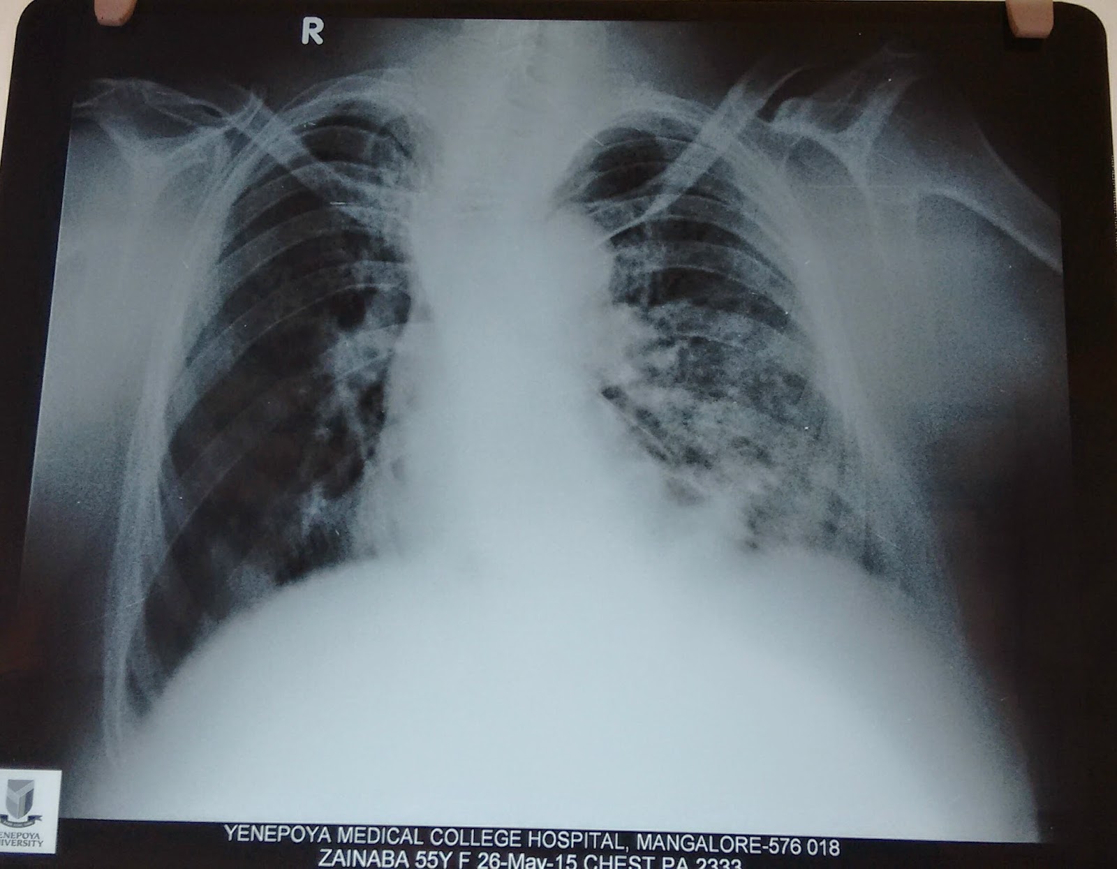
An elderly male presented to us with weakness and giddiness associated with chest pain since a month. Clinical examination revealed an enlarged right supraclavicular node. Rest of the clinical findings were normal.
BUY ONE OF THE BEST BOOKS FOR CHEST RADIOLOGY! PRICES SLASHED WITH INDIAN EDITION


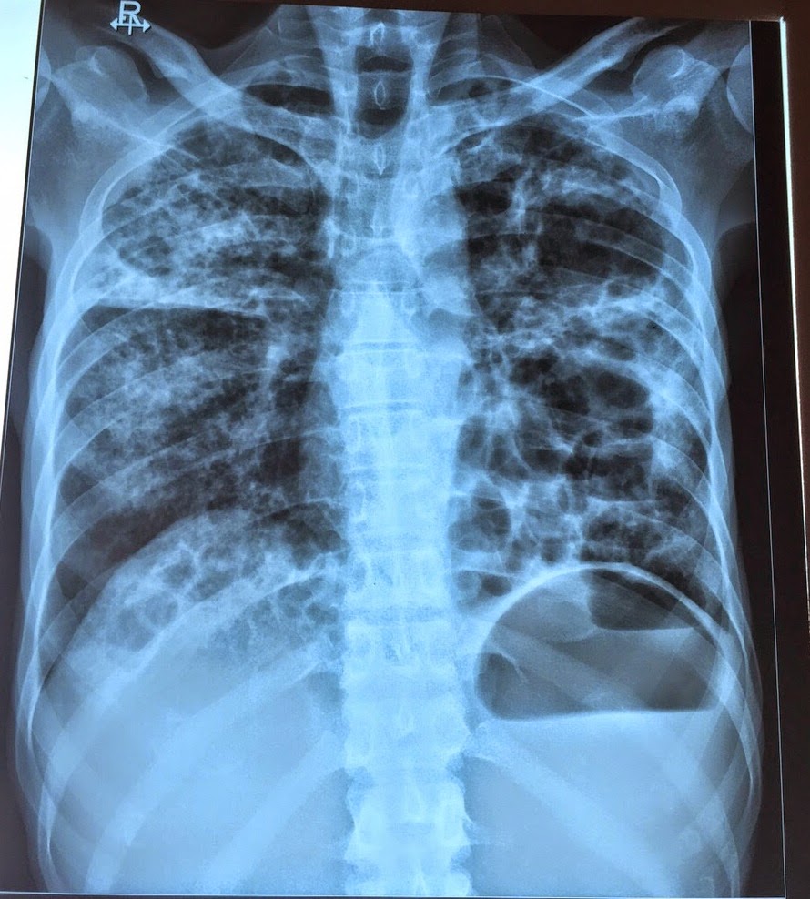
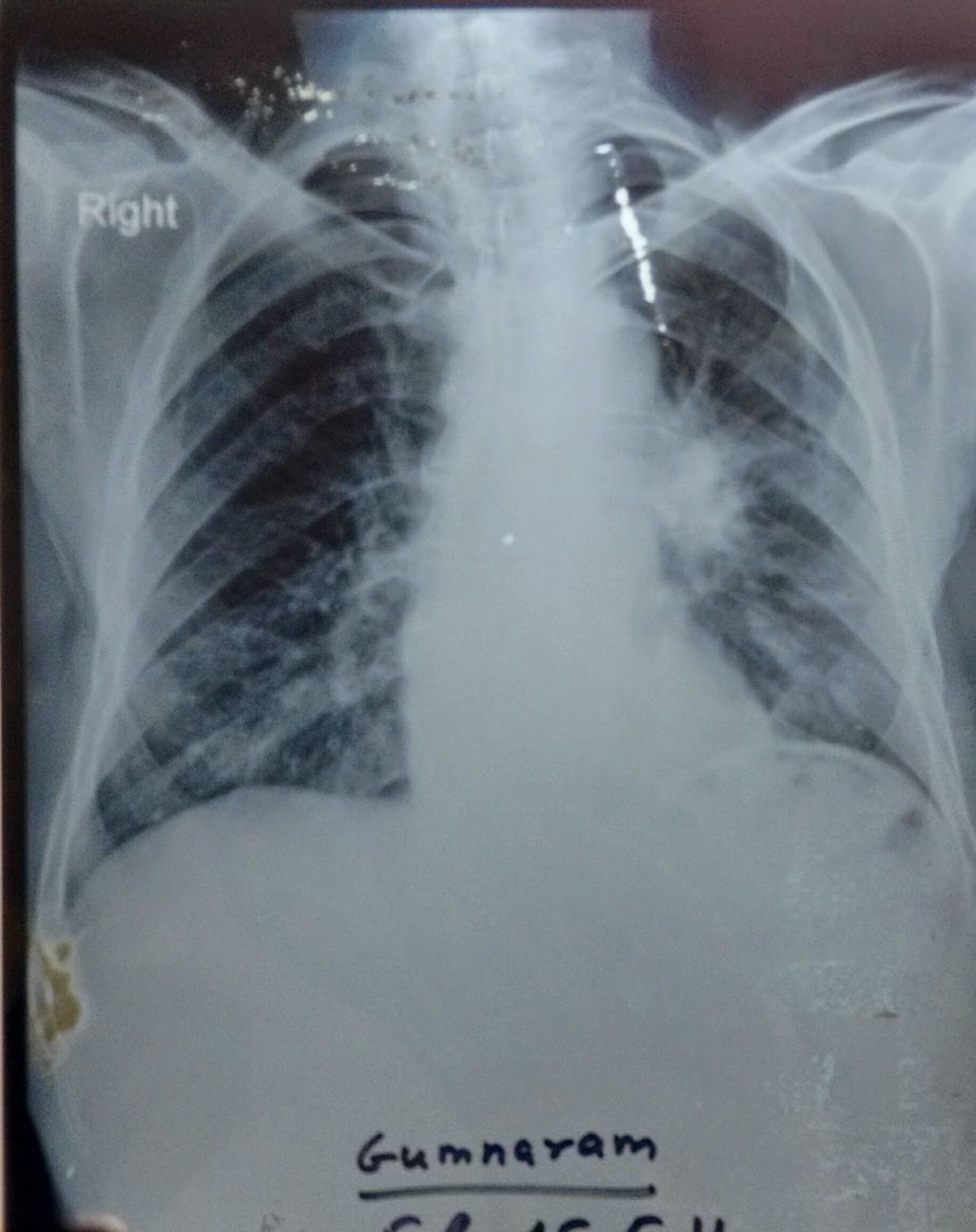
Before proceeding study this chest X Ray carefully.

Chest X ray shows large , well circumscribed, round pulmonary nodules that are distributed in upper mid and lower lung zones bilaterally, some forming into a mass but most of them are seen concentrated in the lower zones and mostly along the peripheral lung fields. also there is a homogenous mass in the left mid zone just lateral to the descending aorta. The diagnosis could be cannon ball secondaries with unknown primary.
Study the CT images before proceeding

The CT scan above shows multiple nodules of varying sizes, concentrated mostly in the lower lobes and left upper lobe bronchus seems occluded partially by the mass. Here study this mediastinal window and coronal cuts

The mediastinal window shows a few lymph nodes in paratracheal and subcarinal area.
We screened his brain , GIT, bones and Genitourinary system for primary Tumor, the scans were normal , further we considered bronchoscopy and Scopy showed a fungating mass in the left upper lobe, Biopsy reports are awaited. Will update once it becomes available.
Cannonball metastases are large, well circumscribed, round pulmonary metastases that appear, well, like cannonballs. 
 
The French term "envolĂ©e de ballons" which translates to "balloons release" is also used to describe this same appearance. 

Causes of Cannon Ball Metastais
Classical- renal cell carcinoma  choriocarcinoma,
                                           
Less commonly - prostate cancer, synovial sarcoma or endometrial carcinoma.
Pulmonary metastases are very common and it is  due to metastatic spread to the lungs from a variety of tumour. It can spread via blood or lymphatics. 
Clinical presentation
Pulmonary metastases are asymptomatic usually,
The constitutional symptoms are related to primary cancerous metastatic condition
Those attributable to the primary tumour dominating. 
Haemoptysis , Chest pain , difficulty in breathing and pneumothorax are sometimes the presenting symptom.
Pathology
Tumour cells reach the lungs via the pulmonary circulation, where they lodge in small distal vessels. 
The most common primaries to result in pulmonary metastases include: 
breast carcinoma
colorectal carcinoma
renal cell carcinoma
uterine leiomyosarcoma
head and neck squamous cell carcinoma
Primaries which most frequently metastasise to lungs (although in themselves much less common tumours) include:
choriocarcinoma
Ewing sarcoma
malignant melanoma
osteosarcoma
testicular tumours
thyroid carcinoma
Radiographic features
Pulmonary metastases characteristically appear as peripheral, rounded nodules of variable size, scattered throughout both lungs . uncommon features include consolidation, cavitation, calcification, haemorrhage and secondary pneumothorax.
HERE IS A SMALL BUT QUITE INFORMATIVE BOOK ON CHEST X RAY DOCTORS SHOULD READ.


Plain radiograph
Plain films are insensitive, although frequently able to make the diagnosis, as often pulmonary metastases are large and numerous.
CT
CT is excellent at visualising pulmonary nodules. Typically metastases appear of soft tissue attenuation, well circumscribed rounded lesions, more often in the periphery of the lung. They are usually of variable size, a feature which is of some use in distinguishing them from a granuloma.
A prominent pulmonary vessel has frequently been noted heading into a metastasis. This is termed the feeding vessel sign.
Some tumours have a predilection for innumerable small metastases (miliary pattern):
Malignant melanoma
Osteosarcoma
Renal cell carcinoma
Thyroid carcinoma
Trophoblastic disease( choriocarcinoma)
Pulmonary metastasis may be single. Seen most frequently  in colorectal carcinoma.
Other primaries which often present with solitary metastases include:
Malignant melanoma
skeletal sarcoma
adenocarcinomas in general
Adenocarcinoma metastases may rather than displace or destroy adjacent lung parenchyma, cells grow in a lepidic fashion (spread along aleveolar walls) resulting in pneumonia-like consolidation. Air bronchograms may also be visible.
Cavitation is present in ~4% of cases. The most common primary is squamous cell carcinoma, most often from the head and neck or from the lung. Other primaries include adenocarcinomas, and sarcomas.
Calcification, although uncommon and more frequently a feature of benign aetiology (e.g. granuloma or hamartoma) is also seen with metastases, particularly those from papillary thyroid carcinoma and adenocarcinomas. Treated metastases, osteosarcomas and chondrosarcomas may also contain calcific densities.
halo of ground-glass opacity representing haemorrhage can be seen, particularly surrounding haemorrhagic pulmonary metastases, such as choriocarcinoma and angiosarcoma.
Treatment and prognosis
In general presence of pulmonary metastases is an ominous finding, indicating poor prognosis. The specific prognosis will however depend on the primary tumour.
Complications

Tumours with prominent necrosis located near a pleural surface may result in a pneumothorax. Osteosarcoma is classically described as the pulmonary metastasis that results in pneumothorax. Another cause of pneumothoraces include cystic or cavitatory pulmonary metastases.

Grand Rounds - Opaque hemithorax.

Pulmonary Medicine Blog By Dr Deepu

Grand Rounds:

This patient an elderly lady presented to us with breathlessness and cough eith sputum with increased sputum in right lateral position. Spo2 was 89% 
Examination revealed trachea deviated to left. Apicak impulse felt in left axilla 5th Ics. Breath sounds diminished on left with added crepitations on left side.
This X ray was taken in emergency room.

 CT confirmed fibrosis of left lung.
Complete white out(opacification) of the hemithorax on CXR has a limited number of causes.
 The differential diagnosis can be zeroed on with one simple observation - the position of the trachea. 
Is it central, pulled or pushed from the side of opacification?
  • pulled trachea : pneumonectomy, total lung collapse, pulmonary fibrosis,pulmonary agenesis
  • central: consolidation, mesothelioma, collapse with effusion. Lung mass 
  • pushed: pleural effusion, diaphragmatic hernia.

Radiology- chest X Ray Spotters.

Pulmonary Medicine Blog By Dr Deepu

spotter 1.
A middle Aged male who is HIV+ presents with a cough of 3 months and cachexia. Auscultation reveals crepitations b/l . Differentials???


Clinical Case - Give Your Diagnosis!!!

Pulmonary Medicine Blog By Dr Deepu
An elderly female came to the outpatient department with a history of cough since 2 weeks minimally productive sputum, she also give history of increased breathlessness since 3 weeks, the symptom of breathlessness being present since three years, she also complains of decreased sleep due to productive cough, and a known hypertensive since 5 years.
  Clinical examination reveals pitting pedal edema and bilateral basal crepitations and no other significant clinical findings were present.
Investigations revealed a total count of 13000 and this chest x ray. EKG was normal. What could be the differential diagnosis????

Spotter : Identify the radiological sign in chest X ray.

Spotter for you: What is your Diagnosis?? What are the differentials and source of such condition.

Chest X Ray- The Diaphragm is unique and provides clue to your diagnosis!!!

Pulmonary Medicine Blog By Dr Deepu

There are a few things which beginners often miss in a chest x ray, one among those is failure to comment on the diaphragms.
Today I am going to discuss importance of tracing diaphragm in a chest X ray with an example.
          Normal diaphragm in a chest X ray has the following characteristics
1.     Trace the diaphragm on right and left
2.     The right diaphragm is usually placed between the fifth and the sixth Rib in the mid clavicular line, It can be seen upto middle of sixth and seventh rib.
3.     The Diaphragms are usually not at the same level on the frontal , erect , inspiratory chest X rays, but they are usually within one rib intercostals space height ( roughly 2 cm) of each other.
4.     The left diaphragm is usually lower than right.
5.     The costophrenic angles should be sharp, making an acute angle.
6.      If the left hemidiaphragm is equal to Right or higher than Right or Right diaphragm is higher than left by more than 3 cms, Causes of diaphragmatic elevation should be considered.
The causes of elevated hemidiaphragm are
1.            Causes above the diaphragm- decreased lung volume due to Lung Collapse, lobectomy, pneumonectomy , fibrosis and pulmonary Hypoplasia
2.            Causes in the diaphragm- Phrenic nerve palsy , diaphragmatic evantration
3.            Causes below the diaphragm- abdominal malignancy, subphrenic abscess, distended   hollow viscus.
After knowing the cause I want to discuss with you a chest x ray where the subtle change in the diaphragm was missed.
Before we proceed Read the chest X ray

 The Chest X ray showed a subtle change in Diaphragm
1. Both the diaphragms are at the same levels.
2. The air shadow underneath the left diaphragm is more prominent.
3. The patient was not evaluated further because chest X Ray appeared normal and sent home with conservative treatment for COPD. 
He came back to our center with hemoptysis one month later referred from the center which treated him initially, a second Radiograph was performed. study the Chest X Ray before proceeding further.  


The chest X ray  now shows features of full blown disease, the hilum is prominent with CORONA RADIATA SIGN suggestive of bronchogenic carcinoma, The left Diaphragm is now placed higher compared to right. Further HR and CECT revealed a tumor in the Left Main bronchus with lymph node metastasis. With Bronchoscopy the diagnosis of squamous cell carcinoma was made.

With this I will end this post, requesting everyone to look at any subtle changes in diaphragm which if ignored may cause some grave diagnosis at a later date.

NEW TREATMENT CAN CURE ASTHMA COMPLETELY!!!!

Pulmonary Medicine Blog By Dr Deepu

Scientists discover asthma's potential root cause and a novel treatment
Cardiff scientists have for the first time identified the potential root cause of asthma and an existing drug that offers a new treatment.

Published today in Science Translational Medicine journal, University researchers, working in collaboration with scientists at King's College London and the Mayo Clinic (USA), describe the previously unproven role of the calcium sensing receptor (CaSR) in causing asthma, a disease which affects 300 million people worldwide.

The team used mouse models of asthma and human airway tissue from asthmatic and non-asthmatic people to reach their findings.

Crucially, the paper highlights the effectiveness of a class of drugs known as calcilytics in manipulating CaSR to reverse all symptoms associated with the condition. These symptoms include airway narrowing, airway twitchiness and inflammation - all of which contribute to increased breathing difficulty.

"Our findings are incredibly exciting," said the principal investigator, Professor Daniela Riccardi, from the School of Biosciences. "For the first time we have found a link airways inflammation, which can be caused by environmental triggers - such as allergens, cigarette smoke and car fumes – and airways twitchiness in allergic asthma.

"Our paper shows how these triggers release chemicals that activate CaSR in airway tissue and drive asthma symptoms like airway twitchiness, inflammation, and narrowing. Using calcilytics, nebulized directly into the lungs, we show that it is possible to deactivate CaSR and prevent all of these symptoms."

Dr Samantha Walker, Director of Research and Policy at Asthma UK, who helped fund the research, said:

"This hugely exciting discovery enables us, for the first time, to tackle the underlying causes of asthma symptoms. Five per cent of people with asthma don't respond to current treatments so research breakthroughs could be life changing for hundreds of thousands of people.

"If this research proves successful we may be just a few years away from a new treatment for asthma, and we urgently need further investment to take it further through clinical trials. Asthma research is chronically underfunded; there have only been a handful of new treatments developed in the last 50 years so the importance of investment in research like this is absolutely essential."
While asthma is well controlled in some people, around one-in-twelve patients respond poorly to current treatments. This significant minority accounts for around 90% of healthcare costs associated with the condition.

According to Cardiff Professor Paul Kemp, who co-authored the study, the identification of CaSR in airway tissue means that the potential for treatment of other inflammatory lung diseases beyond asthma is immense. These include chronic obstructive pulmonary disease (COPD) and chronic bronchitis, for which currently there exists no cure. It is predicted that by 2020 these diseases will be the third biggest killers worldwide.Professor Riccardi and her collaborators are now seeking funding to determine the efficacy of calcilytic drugs in treating asthmas that are especially difficult to treat, particularly steroid-resistant and influenza-exacerbated asthma, and to test these drugs in patients with asthma.
Calcilytics were first developed for the treatment of osteoporosis around 15 years ago with the aim of strengthening deteriorating bone by targeting CaSR to induce the release of an anabolic hormone. Although clinically safe and well tolerated in people, calcilytics proved unsuccessful in treating osteoporosis.
But this latest breakthrough has provided researchers with the unique opportunity to re-purpose these drugs, potentially accelerating the time it takes for them to be approved for use asthma patients. Once funding has been secured, the group aim to be trialling the drugs on humans within two years.
"If we can prove that calcilytics are safe when administered directly to the lung in people, then in five years we could be in a position to treat patients and potentially stop asthma from happening in the first place," added Professor Riccardi.
The study was part-funded by Asthma UK, the Cardiff Partnership Fund and a BBSRC 'Sparking Impact' award.

JOURNALS IN RESPIRATORY SYSTEM / LUNG DISEASES


ECG changes in Acute severe Asthma

Pulmonary Medicine Blog By Dr Deepu

Reversible ECG changes in Acute severe Asthma

Dear Friends I got a call from the emergency department to treat a patient who was gasping for air, the intern at the ER department informed me that the patient had RAD, RBBB and P pulmonale, After examining the patient and taking proper history from the attendants we came to know that the patient is an asthmatic and this was an acute asthmatic attack. Then the intern was baffled by the ECG changes, he thought the event to be a acute cardiac event, I then explained him the ECG changes which are seen in acute asthma. I thought to share the same with you
Here are the ECG changes in Acute Severe Asthma
1.     Sinus tachycardia
2.      Right axis deviation
3.      P pulmonale 
4.      Precordial leads -  voltage of the "p" wave is reduced
5.      Poor progression of the R wave in the precordial leads and marked persistence of the S wave in the left precordial leads
6.      Right bundle branch block
7.      Ventricular premature complexes
8.     Atrial enlargement
9.     Transient ST-segment depression or elevation     in inferior leads ; T-wave abnormalities
10.                        Ventricular repolarization shows a lowered J point with an upward oblique ST segment in the peripheral leads
The mechanism of these electrocardiographic changes appears to depend on the vertical position of the heart caused by over expansion of the lungs and pulmonary arterial hypertension
What are the causes of ECG changes???
1.     Adrenergic stimulation
2.     Hyperventilation
3.     Hyperinflation
4.     Primary or secondary coronary insufficiency
5.     Severity of ECG signs correlates with the degree of airway obstruction.

So, Various ECG changes can occur in acute severe asthma which are nonspecific and these may mimic an acute cardiac event and can cause diagnostic dilemma, most of these changes are reversible usually within 10 days of treatment

The HRCT findings of Bronchiectasis.

Pulmonary Medicine Blog By Dr Deepu


CT findings in bronchiectasis include the following:

·        Let me first describe the specific findings in the bronchiectasis
      Has parallel tram track lines, or
       It may have a signet-ring appearance
     Let me make it more clearer, it is composed of a dilated bronchus  cut in a horizontal section representing the golden ring; with an adjacent  pulmonary artery representing the stone of the Ring. Take a look at the signet ring to get the idea.

·         The diameter of the bronchus lumen is normally 1-1.5 times that of the adjacent vessel; a diameter greater than 1.5 times that of the adjacent vessel suggests bronchiectasis. Let us correlate it in the CT scan
Second Sign is lack of bronchial tapering, Normally as we move to the peripheral parts of the HRCT, the Bronchus should start tapering, as the diameter of the bronchus progressively decreases, whereas in the patients with bronchiectasis this tapering is not seen as the bronchial wall is destroyed and dilated , see the images below
NORMAL BRONCHIAL TAPERING
LACK OF BRONCHIAL TAPERING

 We can also see the abnormal bronchial contour due to the tractional forces applied by the fibrosed and diseased lungs
Visibility of peripheral airways within 1 cm of the pleura
Normally airways are not seen upto 1 cm from the pleura but with bronchiectasis we may be able to see the dilated peripheral airways
ARROWS POINTING TO THE DILATED PERIPHERAL AIRWAYS WITHIN 1 CM OF PLEURA

Its time to describe the non specific findings in the bronchiectasis
Peribronchial cuffing (thickened hazy bronchial wall).
Finger in glove opacities (mucus filled bronchi).
  •Multiple air fluid levels (fluid filled bronchi). 

Peripheral cuffing- here we have a thickened bronchial wall due to constant underlying inflammatory process in the bronchial wall
PATIENT WITH LONG STANDING ABPA SHOWING BRONCHIECTASIS IN THE RIGHT LUNG, WITH BRONCHIAL WALL THICKENING
 Finger in glove opacities( Mucus filled bronchi)
mucus plugging of the bronchus causes bronchus to appear as a gloved hand 

let us see how it appears in the CT Scan


Does this look like a gloved finger???

Next sign is multiple air fluid levels
tThis occurs due to the dilated bronchus and the fluid collected in the dilated bronchus.

Along with these specific and non specific finding we will be able to see few ancillary findings associated with bronchiectasis
Mosaic perfusion.
Air trapping.
Tree in bud opacities.

Mosaic Perfusion
It occurs due to areas alternating areas of normal lung and trapped air in the lungs


Air Trapping, to see air trapping specifically ask for expiratory film, the air gets trapped in the blocked small airways causing dark areas,  whereas the air  is squeezed out from normal lung.

 Tree in Bud Appearance-
occurs due to active infection, study the image and find tree in bud, I have marked it in 2nd image
Image shows bronchiectasis and tree in Bud

tIf you liked the post please comment , comments activates the search engine, Thanks 
Suggested Reading

The Rings !!!The Trams!!!, Chest X Ray Findings in Bronchiectasis


My Next post will be on "SOLITARY PULMONARY NODULE"