Showing posts with label NOTES. Show all posts

PLEURAL EFFUSION

Fluid accumulation in pleural space.
Clinical Picture
  • Patients present with shortness of breath and cough.
  • Can be asymptomatic and recognized on routine CXR.
  • Dullness with decreased breath sounds and mediastinal shift can be seen.
  • Chest x-ray shows fluid in pleural space with classical meniscus. Additional findings will depend on the etiology.
Etiology
Multiple etiologies can give rise to pleural effusion. Following are the common.
  • Congestive heart failure
  • Malignancy
  • Parapneumonic
  • Tuberculosis
  • Rheumatoid arthritis
Pathophysiology
Mechanisms leading to accumulation of fluid in pleural spaces vary. Following are some.
  • Increased hydrostatic pressure eg CHF.
  • Increased capillary permeability eg malignancy
  • Direct extravasation eg Chylothorax
  • Negative pressure induced eg trapped lung
Diagnosis
  • Pleural effusions are categorized as exudates and transudates.
  • Thoracentesis is essential to obtain fluid for appropriate studies based on the clinical setting.
  • Malignancy is the most common cause of exudative effusions and should be ruled out with cytological exam.
  • In undiagnosed effusions one have to consider pleural biopsy either by blind method or by thoracoscopy.
Treatment

  • Appropriate specific therapy based on the etiology.
  • Therapeutic thoracentesis may be required to relieve shortness of breath.
  • Pleural sclerosis will be a consideration in malignant effusions to prevent recurrence.

LUNG METASTASIS

LUNG METASTASIS
Lung is a common site for metastasis from malignancies from other organs.
  • Lung is a capillary bed and the entire cardiac output passes through it, thus it is no surprise tumors get trapped in lungs.
  • Various patterns of metastasis should be recognized. Common patterns are
    • Solitary
    • Cannon balls
    • Lymphangitic
    • Pleural effusions
Clinical Presentation
Mode of clinical presentation varies depending on the pattern of metastasis.
  • Asymptomatic, detected on routine CXR.
  • Cough, hemoptysis, pneumonia, wheezing with endobronchial mets.
  • Shortness of breath, cough with lymphatic spread.
  • Pleuritic pain, cough and shortness of breath with pleural effusions.
Diagnosis
Again depends on the metastatic pattern.
  • FNAB for solitary or multiple lung mets.
  • Sputum cytology and bronchoscopy for endobronchial mets.
  • Pleural fluid cytology for effusions.
  • Tranbronchial lung biopsy for lymphatic spread.
Treatment
Very important treatment issues revolve around the presence and pattern of metastasis.
  • Lung metastasis most often would preclude a surgical curative resection of the primary.
  • Surgical resection of a solitary lung metastasis along with resection of the primary can accomplish cure in certain tumors.
  • Brachytherapy therapy may be useful to relieve symptoms with endobronchial mets.
  • Pleural sclerosis is necessary in pleural effusions.
  • Certain chemotherapeutic agents can sequester in effusions.


MESOTHELIOMA

A mesothelioma is a primary malignant neoplasm of pleura.
Etiology
  • It occurs in patients with prior exposure to asbestos.
  • It has no correlation with cigarette smoking.
Pathology
  • The neoplasm grows to encase the lung and chest wall and spreads locally.
  • Microscopically, the neoplastic cells may resemble mesenchymal stroma (sarcoma) or appear like epithelial cells.
Clinical Features
  • Patients present with SOB, cough, weight loss and chest pain.
  • Pleural effusion is a common mode of presentation.
  • Chest CT reveals pleural effusion and characteristic encasing of lung with tumor mass.
    • CXR
    • CT
  • Diagnosis is difficult with pleural fluid cytology and pleural biopsy.
  • Special stains and an experienced Pathologist are often required for diagnosis.
  • The tumor tends to grow along needle tracks. This is one reason to avoid repeated thoracentesis.
Therapy

  • No therapy has a proven benefit in prolonging life.
  • Patients usually expire within 1-2 years.
  • Extrapleural pneumonectomy is recommended by some centers.

LUNG ABSCESS

LUNG ABSCESS
Definition

Lung abscess is a localized area of liquefactive necrosis of the lung. This would then include necrotizing gram negative and gram positive pneumonias eg. Klebsiella, Staph, Pseudomonas etc. However, by convention we reserve the term lung abscess for necrotizing anaerobic pneumonia.

Prerequisites and Predisposing Conditions
  • Aspiration of a Large Bacterial Inoculum:
    The aspiration of oropharyngeal contents with a bacterial bolus inoculum is the prerequisite for development of lung abscess.
  • Loss of Cough Reflex:
    If the cough reflex is intact, significant aspiration is not possible unless it is overwhelming. Altered sensorium is the most common state when cough reflex is suppressed, thus CVA, drug overdose, alcoholism, post-op state or coma from any cause is the most common predisposing factor for lung abscess.
  • Trouble with Deglutition:
    This occurs with neurological disorders and esophageal diseases. Aspiration to lungs is frequent in this situation even if the cough reflex is intact. In many of the Esophageal diseases the mode of presentation is Lung abscess.
  • Post Obstructive Pneumonia:
    Lung abscess can occur as a complication of post obstructive pneumonia as seen in some patients with lung cancer or foreign body aspiration.
·         Common Segments
·        
The superior segments of RLL, LLL and axillary subsegments of anterior and posterior segments of RUL are common sites for aspiration and will account for 85% of all Lung abscesses.
·         Gravitational forces determine the site of aspiration. Position of the patient at the time of aspiration determines the segment the aspiration is most likely to occur.
·         Basal segments of RLL used to be the most common site for aspiration during 1940 to 1960. During this period ENT surgery and Dental work was done in sitting position with Ether as the anaesthetic. The Right main bronchus is in straight line with Trachea while left main takes of at an angle. In this position gravity facilitates lodging of the aspirate to basal segments of RLL.
·         In supine position and with the patient on back superior segment of RLL is the most dependent segment.
·         In right lateral decubitus position the axillary subsegments of anterior and posterior segments of RUL is the dependant site for aspiration. Abscess is located in the middle of lateral CXR corresponding to RUL bronchus take off.
·         When the patient is on abdomen, aspiration does not occur, thus it is extremely unlikely for any anterior segments, middle lobe and lingula to be the site for aspiration lung abscess. When lung abscess is encountered in these sites on should suspect partial airway obstruction or trouble with deglutition as the predisposing factor for lung abscess.
Clinical Picture
  • Most of the patients present with subacute onset of illness and do not seek medical attention for three to four weeks since the onset of illness.
  • Patients complain of cough, low grade fever, anorexia and weight loss of few weeks duration .
  • Patients often have cough with large amounts of foul smelling sputum.
  • Lack of foul smell does not exclude lung abscess, as 50% of anaerobic infections do not produce a foul smell.
  • The superior segment of RLL , LLL and axillary sub-segments of anterior and posterior segments of RUL usually account for 85% of all aspiration lung abscesses. The lesions will correspond to these sites in CXR.
  • In the early stages one sees consolidation.
  • The inflammatory mass eventually necroses and the necrotic material is expectorated through bronchus.
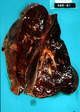
  • The cavity that results has thick wall with irregular lumen. You may note stalactites and stalagmites in the lumen.
  • Air fluid level is the hall mark of Lung abscess.
  • The appearance of the cavity is similar to necrotizing squamous cell cancer of lung and has to be differentiated from it.
·         Bacteriology

Common pathogens are gram positive anaerobes such as peptococci and peptostreptococci, the micro-aerophilic streptococci which are part of normal oropharyngeal flora gram negative anaerobes such as the prevotella (P. Melaninogenicus), Fusobacteria (necrophorum and nucleatum)
Method of Obtaining Specimen
The options are as follows:
  • Sputum Gram Stain:
    May occasionally be helpful if there is a large number of white blood cells and bacteria consistent with oropharyngeal flora.
  • Bronchoscopy:
1.     Triple lumen catheter: Routine aspirates during bronchoscopy is useless for anaerobic cultures. The bronchoscope passes through oropharynx and will be contaminated by the oropharyngeal flora. You need to use triple lumen catheter to avoid contamination and obtain material selectively from the involved segment.
2.     Bronchial lavage: The second option is to obtain a bronchial lavage from the involved segment and perform quantitative bacterial cultures.
  • Fine Needle Aspiration:
    In the pre-antibiotic era needle aspirations of lung abscess were fraught with fear of development of bronchopleural fistula and empyema. With the current option of FNAB under CT guidance, it is being done with increasing frequency and safety. Uncontaminated aspirate can be obtained by this method for cultures. This procedure is often the method of choice for obtaining the specimen in children as other options are not easily feasible in this population.
  • No Need for Cultures:
    When the patient has foul smelling sputum the anaerobic infection is obvious and there may not be a need for confirmation, as many of these procedures are expensive and attendant with some risks. Most of the lung abscess respond to empiric therapy. The primary purpose for culture is to obtain antibiotic sensitivity and can be reserved to cases not responding to empiric therapy.
·         Antibiotic of Choice
·        
Traditionally, penicillin alone was used and produced satisfactory results. Of late, there has been increasing incidence of penicillin resistance in oropharyngeal anaerobes. Hence, penicillin alone is no longer recommended. Metronidazole alone has failed despite its superb anaerobic spectrum due to lack of activity against microaerophilic streptococci which are significant pathogens in lung abscesses. Penicillin added to metronidazole is an acceptable alternative.
·         Clindamycin is the most popular antimicrobial for treatment of lung abscesses and has produced excellent results. The intracellular uptake of clindamycin and its stability in abscess which have low pH and poor vascularity may offer an advantage.
·         Other beta lactams such as ampicillin and sulbactam, ticarcillin or amoxicillin with clavulanate, piperacillin with tazobactam, cefoxitin and cefotetan also have excellent activity against anaerobes and offer expensive alternatives. Imipenem also has excellent activity against anaerobes. Presently, available quinolonoses such as ciprofloxacin, norfloxacin, oflaxacin, etc. have very poor activity against anaerobes and streptococci. Prolonged treatment over several weeks is typically required.
Methods of Drainage
  • Postural Drainage:
    Postural drainage should be instituted in all cases if possible to drain the abscess.This will facilitate drainage of pus. Patient should have empty stomach and prepared with bronchodilator and humidification prior to postural drainage.
In most patients antibiotics and postural drainage is sufficient to cure lung abscess. However in some patients additional drainage options have to be entertained.
  • Bronchoscopy:
    Fiberoptic bronchoscopy is very useful to drain the lung abscess, however cannot be used as a method of drainage daily. This method has to be reserved for situations when the postural drainage and antibiotics are failing to control infection and the cavity is enlarging.
  • Percutaneous Chest Tube:
1.     CT Guided Percutaneous Drainage: One can select the site where the parietal and visceral pleura are adherent and the drain can be placed into the cavity.
2.     In past chest tube was placed into the cavity in two surgical steps. First pleural space was marsupialized by sutures between visceral and parietal pleura. Few days later chest tube was placed into the cavity through this site. This precaution was taken to avoid spilling of pus into pleural cavity .
Role of Bronchoscopy
  • Diagnosis:
    In the past bronchoscopy was done routinely in all patients with lung abscess with the intent of detecting foreign body or cancer. In most the bronchoscopy was non-contributory. Lung abscess is due to aspiration of bacterial bolus and not due aspiration of large foreign body. Cancer and foreign body aspiration account only for a small number of cases. Nowadays bronchoscopy is reserved only when the lung abscess is located in atypical segments or is refractory to therapy.
  • Specimen Collection:
    As discussed under specimen collection methods, bronchoscopy is one method by which we can collect specimen for cultures and sensitivity studies.
  • Drainage of Pus:
    Bronchoscopy is also useful to drain lung abscess trans-bronchially in selected cases.
Role of Surgery


In the pre-antibiotic era surgery was the only method of therapy for lung abscess. In the modern era there is very limited role for surgery in patients with lung abscess. Most of the lung abscesses are curable with antibiotics and postural drainage. Massive hemoptysis is often the most common indication for surgery in the modern days. In patients with partial airway obstruction, lung abscess may increase in size even with antibiotics and one may have to resort to drainage procedure or surgical resection.

KYPHOSCOLIOSIS

KYPHOSCOLIOSIS
Kyphoscoliosis is a disorder characterized by progressive deformity of Spine consisting of lateral and posterior curvatures.
Clinical Picture
  • In majority it is of idiopathic etiology.
  • Deformity results in shortening of height.
  • Patients can be asymptomatic.
  • Mobility of chest wall is impaired, the chest wall is stiff and lung volumes are restricted.
  • Hypoventilation can occur due to small tidal volumes and increased dead space ventilation.
  • V/Q mismatch leads to significant hypoxia, and can progress to symptoms of Cor pulmonale.
  • CXR shows significant distortion of spine and thorax.
  • Mediastinal position is altered and tremendous regional variation in intensity of breath sounds can be noted on physical exam.
  • PFT will show a restrictive defect. Hypoxemia is common.
Therapy
  • Milwaukee brace controls moderate deformities.
  • Surgical correction is attempted in some to fix spine and arrest progression of the deformity.
  • Oxygen on long term may be necessary in patients with significant hypoxemia.


IDIOPATHIC PULMONARY FIBROSIS

Idiopathic pulmonary fibrosis is a disease characterized by diffuse interstitial fibrosis of unknown etiology and frequently occurs between the ages of 50-70.
Clinical Picture
  • Patients present with dry cough and shortness of breath and sometimes with fever, arthralgias and Raynaud's phenomenon.
  • Clubbing (40-70%) tachypnea and persistent coarse basal crackles are encountered on physical exam. Often they are using accessory muscles of inspiration for tidal breathing.
  • CXR: Normal to reticulonodular to honeycombing. HRCT may show ground glass appearance or honeycomb cysts.  In Desquamative interstitial pneumonitis alveolar features can be recognized..
  • PFT shows a restrictive and diffusion defects. Patients are hypoxic and desaturate significantly with exercise.
  • In 25-45%, serum cryoglobulins, RF and ANA are present.
  • BAL: Predominance of neutrophils.
Pathology
Desquamative Interstitial pneumonitis(DIP), Usual Interstitial Pneumonitis(UIP), Lymphocytic Interstitial Pneumonitis( LIP) are various subgroups, probably of the same disease process.
a.      Early IPF: Desquamation predominates where alveolar macrophages fill alveolar spaces (desquamative interstitial pneumonitis or DIP).
b.     Late IPF: Inflammation of alveolar walls continues and fibroblast proliferation with collagen formation occurs (usual interstitial pneumonitis or UIP).
Diagnosis
  •  By exclusion, it will be necessary to rule out other known causes for fibrosis like Silicosis or drugs. A long list of conditions can give rise to interstitial fibrosis 
  • Transbronchial lung biopsy  can identify interstitial fibrosis. Usually need open lung biopsy to confirm.
Treatment
  • Therapy is useful in acute stages when there is significant inflammatory process.
  • Once the fibrosis is chronic or if the chest X-ray has been stable for years or if there is honeycombing, therapy is of no benefit.
  • Corticosteroids + immunosupressants (cyclophosphamide or azathioprine); anecdotal reports describing colchicine or methotrexate or cyclosporine.
  • Oxygen is often prescribed for these patients, probably not influencing pulmonary hypertension.
  • In some cases  Plasmapheresis and Lung Transplant are considerations.
  • Transplantation: 50% two year survival with single lung.
Prognosis

  • In the acute progressive form "Hamman-Rich Syndrome" the prognosis is poor.
  • In the chronic form patients can live long with disability.

HYPERSENSITIVITY PNEUMONITIS

HYPERSENSITIVITY PNEUMONITIS
A type III and IV hypersensitivity reaction to microbial spores, animal proteins and chemicals.
a.      Farmer's lung is the prototypic disease caused by a reaction to Micropolyspora faeni.
b.     Fever, chills, dyspnea, leukocytosis may occur 4-6 hours after exposure and eventually resolve; symptoms and signs may recur on re-exposure.
c.      CXR: Acute - normal to reticulonodular pattern; Chronic - progressive fibrosis, honeycombing.
d.     BAL: Predominance of lymphocytes; increased IgG, IgM.
e.      Serum precipitins to offending antigen present.
f.       Pathology: - Interstitial alveolitis with lymphocytes and non-caseating granulomas (nonspecific); foam cells present (nonspecific)
Diagnosis: Compatible clinical picture, BAL with lymphocytes; serum precipitins; (inhalational challenge).

Therapy: Avoidance of continued inhalational exposure to causative antigen; corticosteroids in severe cases.

GOODPASTURE'S SYNDROME

GOODPASTURE'S SYNDROME
Rare disease characterized by intra-alveolar hemorrhage and glomerulonephritis.
Etiology and Pathogenesis
  • A cytotoxic antibody against glomerular and alveolar basement membrane is responsible for the injury.
  • By activating complement, the antibody causes damage to glomerular and alveolar basement membrane.
  • A respiratory viral infection is believed to initiate production of the antibody.
  • Antigenic similarity between kidney and lung basement membrane accounts for the clinical picture.
Pathology
Clinical Features
  • Pulmonary complaints consist of hemoptysis and dyspnea.
  • Hematuria, proteinuria, red cell casts and renal failure are results of glomerulonephritis.
  • Iron deficiency anemia results from pulmonary hemorrhage.
  • Diffuse bilateral alveolar infiltrates in chest x-ray.
  • Hypoxemia and restrictive defect of PFT's.
  • Leniar deposition of IgG and complement along glomerular and alveolocapillary basement membranes, characteristic of type II reaction in lung and kidney biopsies.
  • EM
  • Antiglomerular basement membrane antibody can be detected in serum.
Therapy

  • Steroid in high doses controls pulmonary hemorrhage.
  • Nephrectomy with dialysis is necessary in some patients.
  • Combination of plasmapheresis and immunosuppressive therapy provides the best results.

EMPYEMA

Empyema is defined as accumulation of pus or fluid with demonstrable bacteria in pleural space.
Clinical Picture
  • Patients present with fever, chills, pleuritic chest pain and cough
  • It can be acute , subacute or chronic.
  • Leukocytosis with shift to left and Doehle bodies can be noted on CBC.
  • Besides findings of effusion , clubbing, chest wall erythema and edema, increased warmth may be noted on physical exam.
  • CXR will show effusion and cannot be distinguished from other types. Loculated effusions should raise suspicion for empyema.
  • Lack of fever or leukocytosis does not rule out empyema.
Etiology and Pathophysiology
  • Empyema most often is due to extension of infection from pneumonia. Staphylococcal, gram negative and anaerobic infections are common infections presenting in this mode.
  • Anaerobic infections can seed pleura and start as the primary site of infection without a preceding pneumonitis.
  • It could also follow contamination of pleural space from non-sterile pleural taps.
Diagnosis
  • Pleural tap should be done immediately once empyema is a consideration. If the fluid is grossly purulentdiagnosis is established.
  • Gram stain of the pleural fluid and cultures for aerobes and anaerobes should be obtained.
  • If the fluid is not purulent then obtain Ph, glucose and LDH. This will help categorize parapneumonic effusions as simple and complicated effusions.
  • CBC and cultures of sputum and blood are routine.
Treatment

  • Empyema should be drained immediately with chest tube insertion..
  • Appropriate Antibiotics should be started immediately, empiric to start with followed by specific drug based on culture.
  • Streptokinase is useful to break up adhesions if there are loculations.
  • Some patients not responding to this regimen may require thoracotomy to lyse adhesions . This can be accomplished by thoracoscope. Some would require decortication, if a thick pyemic peel has formed and prevent lung expansion.

DRUG INDUCED LUNG DISEASE

DRUG INDUCED LUNG DISEASE
Adverse drug reactions account for 5% of hospital admissions and occur in 18% of hospitalized patients.
Bleomycin
1.     Causes an interstitial pneumonitis that may lead to fibrosis (10-20% of patients receiving bleomycin.)
a.      Factors which increase the risk of bleomycin-induced pulmonary toxicity.
- Age (>70 yrs), dose, route of administration, radiation therapy, 02 therapy, renal function, other chemotherapeutic agents.
2.     Symptoms include dyspnea, cough, fever.
3.     CXR: Basilar reticular or fine nodular changes.
4.     Diagnosis: Diffuse uptake on gallium scan; neutrophils on BAL; transbronchial biopsy (diagnosis made in setting of compatible clinical, radiologic and/or histologic findings).
5.     Mortality: 1-7%.
6.     Therapy: Reduce Fi02 if possible, trial of corticosteroids.
Amiodarone
1.     Injury mediated by both direct and indirect mechanisms.
a.      Greater risk of toxicity with daily maintenance dose of more than 400 mg.
b.     Acute and subacute forms exists; occasionally fulminant course with ARDS.
2.     Symptoms include dyspnea and occasionally cough, fever, and chest pain.
3.     CXR: Diffuse interstitial changes; sometimes upper lobe predilection.
4.     PFT's: Decreased DLCO; reduced volumes.
5.     Histology: Alveolar septal thickening with inflammatory cells, intra-alveolar foam macrophages.
6.     Diagnosis: Usually by exclusion; compatible clinical picture.
7.     Therapy:
a.      Discontinue amiodarone and switch to alternative antiarrhythmic agent
b.     Corticosteroids for severe cases.
Other drugs which may cause interstitial pneumonitis/fibrosis: alkylating agents
1.     Alkylating agents (busulfan, cyclophosphamide, melphalan, chlorambucil)
2.     Nitrosoureas (carmustine, lomustine)
3.     Mitomycin.
Other drugs which may cause hypersensitivity pneumonitis:
1.     Methotrexate
2.     Gold salts
3.     Nitrofurantoin
4.     Sulfasalazine.
Drugs which may cause noncardiac pulmonary edema
1.     Salicylates
2.     Thiazides
3.     Narcotics
4.     Tocolytic agents.
Radiation Therapy
1.     Results in pneumonitis or fibrosis.
a.      Pneumonitis generally appears between 2 to 6 months after radiation; steroids may benefit if given early.

b.     Fibrosis appears between 6 to 12 months after radiation; may progress to respiratory failure; steroids usually not helpful.

DIFFUSE ALVEOLAR HEMORRHAGE

DIFFUSE ALVEOLAR HEMORRHAGE
Diffuse pulmonary hemorrhage is one of the conditions in the differential of acute diffuse alveolar pattern on CXR.
Clinical Picture
  • Patients present with acute onset of shortness of breath and cough.
  • Hemoptysis may or may not be present.
  • Chest x-ray shows diffuse alveolar pulmonary infiltrates.
  • tachypnea with bilateral crackles can be heard on physical exam.
  • Blood gases show hypoxia with widened A-a gradient and alveolar hyperventilation.
Etiology
Multiple etiologies can give rise to pulmonary hemorrhage. Following are the common.
  • Thrombocytopenia
  • Goodpasture's syndrome
  • Wegener's granulomatosis
  • Systemic lupus erythematosis
  • Idiopathic pulmonary hemorrhage
Pathology
Alveolar spaces are filled with blood. Lung is heavy and feels consolidated. rest of the findings will depend on the etiology.
Diagnosis
  • Constellation of hemoptysis, diffuse alveolar infiltrates on CXR, unexplained drop in hemoglobin usually raises suspicion for pulmonary hemorrhage.
  • If PFT can be done, increase in diffusion capacity can be seen due to sequestration of blood in lungs.
  • Bronchoalveolar alveolar lavage will show RBC's and Iron stain is positive in macrophages.
Treatment

  • Supportive care.
  • Appropriate therapy based on the etiology.