The Rings !!!The Trams!!!, Chest X Ray Findings in Bronchiectasis

Pulmonary Medicine Blog By Dr Deepu

  
Bronchiectasis is  an abnormal and permanent distortion of one or more of the conducting bronchi or airways.
In 1950, Reid characterized bronchiectasis as cylindrical, cystic, or varicose types.


Types of bronchiectasis


Cylindrical Bronchiectasis
Mild Form shows Tram Track Appearance

Varicose Bronchiectasis
Moderate Form appears as string of pearls

Cystic/ Saccular Bronchiectasis
Severe Form appears like Bunch of Grapes


       Chest radiography Chest radiography (CXR) is usually the initial study performed in both suspected bronchiectasis and the evaluation of nonspecific respiratory symptoms, such as dyspnoea and haemoptysis, when bronchiectasis may be identified incidentally.

Signs on CXR are the identification of
Read This X Ray Before Proceeding Further
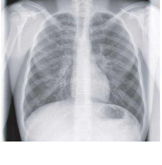

1.     Parallel linear densities, tram-track opacities.

what was seen on the chest X ray, it is nothing but the tram line appearance, unable to spot it, here comes the Modified image
Now Compare the previous X Ray with the one above , Here are few examples of tram line shadows
The black arrows points towards tram line and the white to shadows which will be discussed below
Read this X ray before proceeding
What Can we see here
if you have got it proceed further
What we see here is the ring shadows, there are many other ring shadows in the x ray , only a few are  marked
One More X ray below showing the ring shadows in Cystic Fibrosis Patient 


2.     Ring shadows reflecting thickened and abnormally dilated bronchial walls. These bronchial abnormalities  may vary from subtle or barely perceptible 5-mm ring shadows to obvious cysts.
3.      Fluid or mucous filling of bronchi is seen and leads to Tubular branching opacities conforming to the expected bronchial branching pattern.


4.     The Definition of vessel walls is lost due to  peribronchial fibrosis.
5.     Signs of complications/exacerbations, such as patchy densities due to mucoid impaction and consolidation
6.      Volume loss secondary to bronchial mucoid obstruction or chronic cicatrisation, are also seen.
7.     In the more diffuse forms , such as cystic fibrosis (CF), generalised hyperinflation and oligaemia are often present, consistent with severe small airways obstruction.
The radiograph may raise the initial suspicion of bronchiectasis, triggering more definitive imaging. 
CXR also plays a role in the follow-up of bronchiectasis and management of exacerbations.Although CXR has limitations in specificity in diagnosing bronchiectasis and in detecting early or subtle changes, it is useful for assessing more florid cases of bronchiectasis, in CF and in follow-up of bronchiectatic patients. Computed tomography.

suggested Reading


CASE OF THE WEEK- "Chest Medicine challenge"- case no 03( 24th April- 1st May).

Pulmonary Medicine Blog By Dr Deepu

A 20-year-old woman comes to the ED with a 2-week history of gradually progressive shortness of breath, orthopnea, and dysphagia. She reports an 18-lb (8 kg) weight loss and subjective fevers over the same period of time. She also describes chest tightness and a nonproductive cough. The patient was empirically treated by her private physician with a course of antibiotics without improvement 7 days ago. She was told that there was an abnormality on her chest radiograph and a chest CT scan was performed, but she did not follow up on the results. She is a never-smoker and has no history of asthma. On physical examination, the patient is in mild-to-moderate respiratory distress with a respiratory rate of 32/min; BP, 152/84 mm Hg; and pulse rate of 130/min. Her temperature is 38.4° C. Neck examination reveals jugular venous distention. On lung examination, there is diff use wheezing. Her current posterior-anterior and lateral chest radiographs are shown (Figs 1-A, 1-B). A chest CT scan is ordered, but the patient becomes more short of breath (accentuated when supine in preparation for the CT scan), restless, and confused. Her respiratory rate increases to 40/min, and her oxygen saturation is 88% when breathing 50% oxygen by face mask.

The next step in this patient’s management should be:

A. Awake intubation in the semiupright position.
B. Rapid sequence intubation.
C. Noninvasive mechanical ventilation.
D. Emergency tracheostomy.
also comment on the CT and Chest X Ray

Please come back on Tuesday for answer.

CHEST RADIOLOGY

Air Bronchogram Sign.


Basics Of Chest X Ray Part-6, The Lungs, Pleura And The Chest Wall.

Pulmonary Medicine Blog By Dr Deepu

Lung abnormalities mostly present as areas of increased density, which can be divided into the following patterns:

Consolidation
Atelectasis
Nodule or mass - solitary or multiple
Interstitial
Less frequently areas of decreased density are seen as in emphysema or lungcysts.

BEST BOOK FOR CHEST RADIOLOGY


Chest X-Ray - Lung disease.
Consolidation

Atelectasis


Nodule - Masses

Solitary pulmonary node 
Interstitial pattern


Interstitial lung diseases will be discussed in coming posts.

Pleura

Pleural fluid

It takes about 200-300 ml of fluid before it comes visible on an CXR (figure).
About 5 liters of pleural fluid are present when there is total opacification of the hemithorax.

Total opacification of the right hemithorax in a patient with pleuritis carcinomatosa on both sides.
SUGGESTED BOOKS 


On the right there is only some air visible in the major bronchi creating an air bronchogram within the compressed lung.
Pleural fluid may become encysted.


Here we see fluid entrapped within the fissure.
This can sometimes give the impression of a mass and is called 'vanishing tumor'.

Pneumothorax

The table lists the most common causes of a pneumothorax.
The other cystic lungdisease which causes pneumothorax is Langerhans cell histiocytosis (LCH) which is seen in smokers.
Study the CXR.

There are two important findings.
The retracted visceral pleura is seen (blue arrow) which indicates that there is a pneumothorax.
There is a horizontal line visible (yellow arrow).

Normally there are no straight lines in the human body unless when there is an air-fluid level.
This means that there is a hydro-pneumothorax.
When a pneumothorax is small, this air-fluid level can be the only key to the diagnosis of a pneumothorax.
Study the CXR.
There are 3 important findings.
Notice that the mediastinum is slightly displaced to the left.
Does this mean that there is a tension pneumothorax?
Do you have an idea about the cause of the pneumothorax?
There is a hydropneumothorax.

Notice the air-fluid level (blue arrow).
The upper lobe is still attached to the chest wall by adhesions.
Maybe this patient was treated for a prior pneumothorax.
There is a lung cyst in the upper lobe (red arrow).
So we can assume that the pneumothorax has something to do with a cystic lung disease.
Since this patient is a woman, lymphangioleiomyomatosis (LAM) is a possible diagnosis.
LAM is a rare lung disease that results in a proliferation of smooth muscle throughout the lungs resulting in the obstruction of small airways leading to pulmonary cyst formation and pneumothorax.
LAM also occurs in patients who have tuberous sclerosis.
Study the CXR.

What is your diagnosis?
This is not a pneumothorax but a skin fold.
The radiography was performed supine with a CR cassette inserted underneath the patient, which resulted in a skinfold.
Notice that there are lung markings beyond the apparent pneumothorax.
Here two CXRs of another patient with obvious skinfolds.
Recognition of a pneumothorax depends on the volume of air in the pleural space and the position of the body.
On a supine radiograph a pneumothorax can be subtle and approximately 30% of pneumothoraces are undetected.
A sign to look for is the 'deep sulcus sign'.
It represents lucency of the lateral costophrenic angle extending toward the hypochondrium (Figure).
The image is of a patient in the ICU who is on mechanical ventilation. There was an acute exacerbation of the dyspnea.
There is a deep sulcus sign on the left
Notice that the left hemidiaphragm is depressed.
This is an important finding since it indicates a tension pneumothorax.
The image on the below is after insertion of an intercostal drain.
Notice that the diaphragm has regained its normal appearance.
Pleural opacities
The table lists the most common causes of pleural opacities.

Pleural plaques
The CXR shows multiple opacities.

They have irregular shapes and do not look like a lung masses or consolidations.
Some of these opacities are clearly bordering the chest wall (red arrows).
All these findings indicate that we are dealing asbestos related pleural plaques.
Asbestos related pleural plaques are usually:
bilateral and extensive.
covering the dome of the diaphragm.
Unilateral pleural calcifications are usually due to:

infection (TB)
empyema
hemorrhagic
Pleural hematoma

These images are of a patient, who had a pleural opacity after a chest trauma.
It was believed to be a hematoma and resolved spontaneously.

Chest wall

Ribfractures
The most common identified chest wall abnormalities are old ribfractures.
The CXR shows many rib deformities due to old fractures.

When a rib fracture heals, the callus formation may create a mass-like appearance (blue arrow).

Sometimes a CT is necessary to differentiate a healing fracture from a lung mass.
Notice the large lung volume and the enlarged pulmonary vessels.
Probably we are dealing with pulmonary arterial hypertension in a patient with COPD.
The second most common chest wall abnormalities that we see on a CXR are metastases in vertebral bodies and ribs.

Notice the expansile mass in the posterior rib on the right.

Abdomen

The most obvious finding on this CXR is free air under the diaphragm.
This finding indicates a bowel perforation, unless when the patient had recent abdominal surgery and there is still some air left in the abdomen, which can stay there for several days.
There is another subtle finding in the left upper lobe.
A subtle density projecting over the first rib - hidden area - proved to be a lungcarcinoma.
Here another patient with free abdominal air.

Notice the very thin regular line which is the diaphragm (arrow).

The “BIG RIB SIGN” and “VERTICAL DISPLACEMENT SIGN” In Lateral Chest X Ray.

Pulmonary Medicine Blog By Dr Deepu

The “BIG RIB SIGN” and “VERTICAL DISPLACEMENT SIGN”- To Differentiate The Right and Left Ribs on Lateral Chest Radiographs

Today I would like to share with you two important signs in Chest X Ray- The Big Rib Sign and Vertical Displacement Sign.

Diseases of the ribs and diaphragm are often identified only at lateral chest radiography. Therefore, it is important to use a reliable technique for differentiating the right and left ribs. Two techniques—the “big rib” sign and the “vertical displacement “sign—can be used to localize the right and left ribs at lateral chest radiography. Conventially lateral radiographs are taken with left side near the film. But lateral view can be obtained as Right or left  as per clinicians request, then the principle remains the same but only the magnified sides change .

The big rib sign is a technique that exploits the difference in magnification between the right and left sides on lateral chest radiographs.


The side of the rib cage farther from the film is magnified more than the side closer to the film. On a well-positioned left lateral chest radiograph, the right ribs appear larger than the left ribs . This difference in rib size is more easily detected posteriorly where the x-ray beam is tangential to the ribs but can be appreciated at all corresponding points along the curvature of the two ribs. In addition, rotating the patient may enhance or reduce the magnification effect because, with such rotation, the x-ray beam is transmitted through different portions of the ribs, which are thin medially and thick laterally. In a left lateral projection, when the posterior portions of the right and left ribs appear comparable in size, the hemidiaphragm traceable to the most anterior ribs is the right hemidiaphragm. Otherwise, the significantly larger ribs are the right ribs, which are farther from the film.

The big rib sign is very useful but is not perfect because the magnification difference between the right and left ribs is only 10%. For example, if the width of a rib is 5 mm, the observed difference between sides is only 0.5 mm, which is not always sufficient to enable differentiation of the two sides. Furthermore, the big rib sign is not applicable when the posterior ribs are superimposed.

Another Sign is the vertical displacement sign, which is an easy, reliable, and precise method for differentiating the right and left ribs on lateral radiographs .

 The vertical displacement sign is based primarily on the vertical divergence of the x-ray beam rather than on the magnification of the ribs. Because the right rib cage is farther from the film, the projection of the right ribs on a lateral radiograph will fan out and diverge in a vertical direction to a greater degree than that of the left ribs. Thus, the right side can be distinguished from the left by the vertical displacement of the paired ribs.

The vertical displacement sign is usually applicable even when the posterior ribs are partially superimposed. It is usually possible to identify which rib is higher or lower even when they overlap. The vertical displacement sign can be used as an alternative when the big rib sign is not applicable.

Also Read

Air Bronchogram Sign.


The ‘Dark Bronchus’ Sign


reference:
1. http://pubs.rsna.org/doi/pdf/10.1148/radiographics.19.1.g99ja02105


Air Bronchogram Sign.

Pulmonary Medicine Blog By Dr Deepu

Air bronchogram

An air bronchogram is a tubular outline of an airway made visible by filling of the surrounding alveoli by fluid or inflammatory exudates. It is almost always caused by a pathologic airspace/alveolar process, in which something other than air fills the alveoli.

 Six causes of air bronchograms are
Lung consolidation
Pulmonary edema
Nonobstructive pulmonary atelectasis
Severe interstitial disease
Neoplasm
Normal expiration.
In The Image Air Bronchogram is seen.
The Magnified Image Showing Air Bronchogram On CXR and Confirmed With HRCT


The phenomenon is Characterised by  air-filled bronchi (dark) being made visible by the opacification of surrounding alveoli (grey/white). It is almost always caused by a pathologic airspace/alveolar process, in which something other than air fills the alveoli.
 Air bronchograms will not be visible if the bronchi themselves are opacified (e.g. by fluid) and thus indicate patent proximal airways.
Air bronchograms that persist for weeks despite appropriate antimicrobial therapy should raise the suspicion of a neoplastic process. CT may be planned in such cases.

Also See