Showing posts with label DIFFUSE PULMONARY HEMORRHAGE. Show all posts

WEGENERS GRANULOMATOSIS

WEGENERS GRANULOMATOSIS
  • Rare disease characterized by necrotizing granulomas and vasculitis of upper and lower respiratory tracts; also systemic vasculitis with focal necrotizing glomerulonephritis.
  • Triad of upper respiratory tract, lung and kidney involvement is the classical mode of presentation.
  • Systemic vasculitis may be manifested by skin, eye and joint findings.
  • Limited form with long survival when the disease is predominantly restricted to lungs.
  • Etiology is unknown.
Clinical Picture Diagnosis
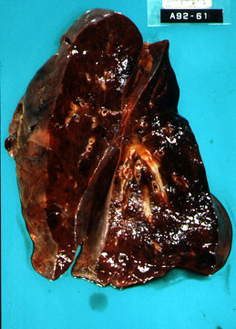
  • Waxing and waning lung lesions, single or multiple often with cavitation.
  • CXR: Bilateral nodules ranging from 1 to 9 cm in size; may also see diffuse interstitial disease and alveolar hemorrhage.
  • Antineutrophilic cytoplasmic antibodies (ANCA) Percutaneous renal biopsy; lung biopsy.
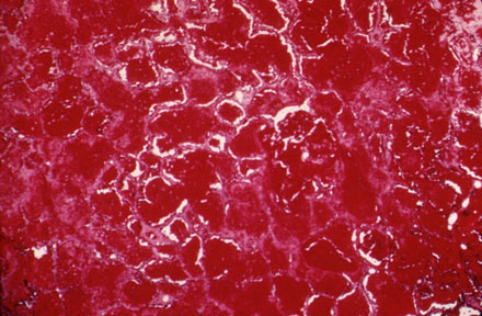
  • Depending on the organ involved, necrotizing nasal lesions, glomerulonephritis, CNS or skin involvement can be seen on biopsy.
Therapy
  • Cytotoxic therapy (Cyclophosphamide) with steroids produce rapid reversal of disease.


GOODPASTURE'S SYNDROME

GOODPASTURE'S SYNDROME
Rare disease characterized by intra-alveolar hemorrhage and glomerulonephritis.
Etiology and Pathogenesis
  • A cytotoxic antibody against glomerular and alveolar basement membrane is responsible for the injury.
  • By activating complement, the antibody causes damage to glomerular and alveolar basement membrane.
  • A respiratory viral infection is believed to initiate production of the antibody.
  • Antigenic similarity between kidney and lung basement membrane accounts for the clinical picture.
Pathology
Clinical Features
  • Pulmonary complaints consist of hemoptysis and dyspnea.
  • Hematuria, proteinuria, red cell casts and renal failure are results of glomerulonephritis.
  • Iron deficiency anemia results from pulmonary hemorrhage.
  • Diffuse bilateral alveolar infiltrates in chest x-ray.
  • Hypoxemia and restrictive defect of PFT's.
  • Leniar deposition of IgG and complement along glomerular and alveolocapillary basement membranes, characteristic of type II reaction in lung and kidney biopsies.
  • EM
  • Antiglomerular basement membrane antibody can be detected in serum.
Therapy

  • Steroid in high doses controls pulmonary hemorrhage.
  • Nephrectomy with dialysis is necessary in some patients.
  • Combination of plasmapheresis and immunosuppressive therapy provides the best results.

DIFFUSE ALVEOLAR HEMORRHAGE

DIFFUSE ALVEOLAR HEMORRHAGE
Diffuse pulmonary hemorrhage is one of the conditions in the differential of acute diffuse alveolar pattern on CXR.
Clinical Picture
  • Patients present with acute onset of shortness of breath and cough.
  • Hemoptysis may or may not be present.
  • Chest x-ray shows diffuse alveolar pulmonary infiltrates.
  • tachypnea with bilateral crackles can be heard on physical exam.
  • Blood gases show hypoxia with widened A-a gradient and alveolar hyperventilation.
Etiology
Multiple etiologies can give rise to pulmonary hemorrhage. Following are the common.
  • Thrombocytopenia
  • Goodpasture's syndrome
  • Wegener's granulomatosis
  • Systemic lupus erythematosis
  • Idiopathic pulmonary hemorrhage
Pathology
Alveolar spaces are filled with blood. Lung is heavy and feels consolidated. rest of the findings will depend on the etiology.
Diagnosis
  • Constellation of hemoptysis, diffuse alveolar infiltrates on CXR, unexplained drop in hemoglobin usually raises suspicion for pulmonary hemorrhage.
  • If PFT can be done, increase in diffusion capacity can be seen due to sequestration of blood in lungs.
  • Bronchoalveolar alveolar lavage will show RBC's and Iron stain is positive in macrophages.
Treatment

  • Supportive care.
  • Appropriate therapy based on the etiology.