Showing posts with label BRONCHIECTASIS. Show all posts

Image Archives- Bronchiectasis

By Dr Deepu

Chest X ray shows bilateral cystic bronchiectasis with air fluid levels, suggesting ongoing infective process

Kartageners Syndrome

By Dr Deepu
See the question
The answer is b."Kartagener syndrome"

Kartagener syndrome is the inheritable disorder of dextrocardia, chronic sinusitis (with the formation of nasal polyps), and bronchiectasis. Patients may also present with situs inversus.The disorder is due to a defect that causes the cilia within the respiratory tract epithelium to become immotile. Cilia of the sperm are also affected

The HRCT findings of Bronchiectasis.

Pulmonary Medicine Blog By Dr Deepu


CT findings in bronchiectasis include the following:

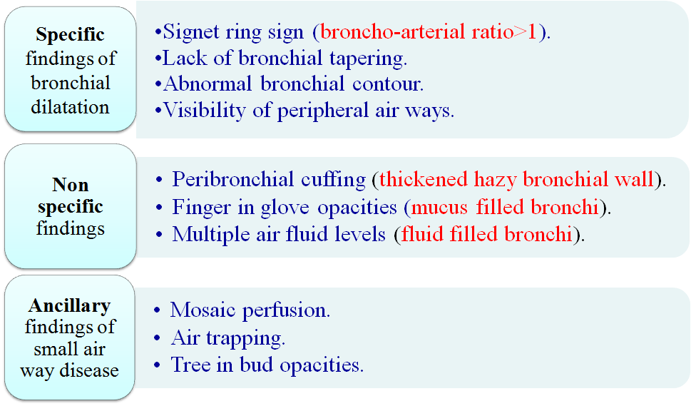
·        Let me first describe the specific findings in the bronchiectasis
      Has parallel tram track lines, or
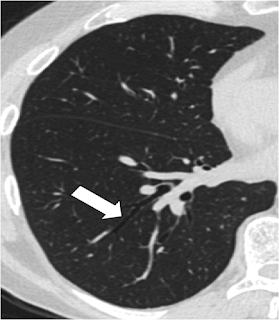
       It may have a signet-ring appearance
     Let me make it more clearer, it is composed of a dilated bronchus  cut in a horizontal section representing the golden ring; with an adjacent  pulmonary artery representing the stone of the Ring. Take a look at the signet ring to get the idea.

·         The diameter of the bronchus lumen is normally 1-1.5 times that of the adjacent vessel; a diameter greater than 1.5 times that of the adjacent vessel suggests bronchiectasis. Let us correlate it in the CT scan
Second Sign is lack of bronchial tapering, Normally as we move to the peripheral parts of the HRCT, the Bronchus should start tapering, as the diameter of the bronchus progressively decreases, whereas in the patients with bronchiectasis this tapering is not seen as the bronchial wall is destroyed and dilated , see the images below
NORMAL BRONCHIAL TAPERING
LACK OF BRONCHIAL TAPERING

 We can also see the abnormal bronchial contour due to the tractional forces applied by the fibrosed and diseased lungs
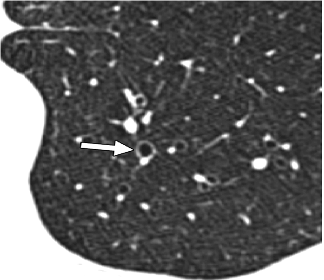
Visibility of peripheral airways within 1 cm of the pleura
Normally airways are not seen upto 1 cm from the pleura but with bronchiectasis we may be able to see the dilated peripheral airways
ARROWS POINTING TO THE DILATED PERIPHERAL AIRWAYS WITHIN 1 CM OF PLEURA

Its time to describe the non specific findings in the bronchiectasis
Peribronchial cuffing (thickened hazy bronchial wall).
Finger in glove opacities (mucus filled bronchi).
  •Multiple air fluid levels (fluid filled bronchi). 

Peripheral cuffing- here we have a thickened bronchial wall due to constant underlying inflammatory process in the bronchial wall
PATIENT WITH LONG STANDING ABPA SHOWING BRONCHIECTASIS IN THE RIGHT LUNG, WITH BRONCHIAL WALL THICKENING
 Finger in glove opacities( Mucus filled bronchi)
mucus plugging of the bronchus causes bronchus to appear as a gloved hand 

let us see how it appears in the CT Scan


Does this look like a gloved finger???

Next sign is multiple air fluid levels
tThis occurs due to the dilated bronchus and the fluid collected in the dilated bronchus.

Along with these specific and non specific finding we will be able to see few ancillary findings associated with bronchiectasis
Mosaic perfusion.
Air trapping.
Tree in bud opacities.

Mosaic Perfusion
It occurs due to areas alternating areas of normal lung and trapped air in the lungs


Air Trapping, to see air trapping specifically ask for expiratory film, the air gets trapped in the blocked small airways causing dark areas,  whereas the air  is squeezed out from normal lung.

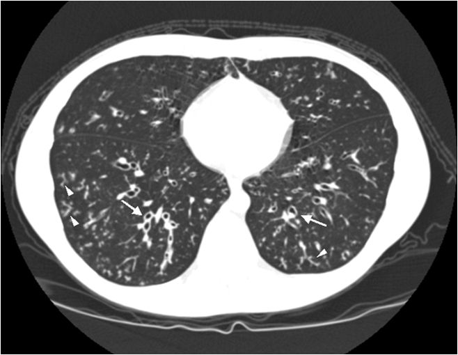
 Tree in Bud Appearance-
occurs due to active infection, study the image and find tree in bud, I have marked it in 2nd image
Image shows bronchiectasis and tree in Bud

tIf you liked the post please comment , comments activates the search engine, Thanks 
Suggested Reading

The Rings !!!The Trams!!!, Chest X Ray Findings in Bronchiectasis


My Next post will be on "SOLITARY PULMONARY NODULE"

The Rings !!!The Trams!!!, Chest X Ray Findings in Bronchiectasis

Pulmonary Medicine Blog By Dr Deepu

  
Bronchiectasis is  an abnormal and permanent distortion of one or more of the conducting bronchi or airways.
In 1950, Reid characterized bronchiectasis as cylindrical, cystic, or varicose types.


Types of bronchiectasis


Cylindrical Bronchiectasis
Mild Form shows Tram Track Appearance

Varicose Bronchiectasis
Moderate Form appears as string of pearls

Cystic/ Saccular Bronchiectasis
Severe Form appears like Bunch of Grapes


       Chest radiography Chest radiography (CXR) is usually the initial study performed in both suspected bronchiectasis and the evaluation of nonspecific respiratory symptoms, such as dyspnoea and haemoptysis, when bronchiectasis may be identified incidentally.

Signs on CXR are the identification of
Read This X Ray Before Proceeding Further


1.     Parallel linear densities, tram-track opacities.

what was seen on the chest X ray, it is nothing but the tram line appearance, unable to spot it, here comes the Modified image
Now Compare the previous X Ray with the one above , Here are few examples of tram line shadows
The black arrows points towards tram line and the white to shadows which will be discussed below
Read this X ray before proceeding
What Can we see here
if you have got it proceed further
What we see here is the ring shadows, there are many other ring shadows in the x ray , only a few are  marked
One More X ray below showing the ring shadows in Cystic Fibrosis Patient 


2.     Ring shadows reflecting thickened and abnormally dilated bronchial walls. These bronchial abnormalities  may vary from subtle or barely perceptible 5-mm ring shadows to obvious cysts.
3.      Fluid or mucous filling of bronchi is seen and leads to Tubular branching opacities conforming to the expected bronchial branching pattern.


4.     The Definition of vessel walls is lost due to  peribronchial fibrosis.
5.     Signs of complications/exacerbations, such as patchy densities due to mucoid impaction and consolidation
6.      Volume loss secondary to bronchial mucoid obstruction or chronic cicatrisation, are also seen.
7.     In the more diffuse forms , such as cystic fibrosis (CF), generalised hyperinflation and oligaemia are often present, consistent with severe small airways obstruction.
The radiograph may raise the initial suspicion of bronchiectasis, triggering more definitive imaging. 
CXR also plays a role in the follow-up of bronchiectasis and management of exacerbations.Although CXR has limitations in specificity in diagnosing bronchiectasis and in detecting early or subtle changes, it is useful for assessing more florid cases of bronchiectasis, in CF and in follow-up of bronchiectatic patients. Computed tomography.

suggested Reading


BRONCHIECTASIS

BRONCIECTASIS
Definition
Bronchiectasis is a severe, chronic infection of the lung causing pronounced permanent abnormal dilatation of bronchi and bronchioles.
Pathology
  • Chronic, necrotizing infection of bronchi in a segment or multiple segments of the lung.
  • Infection damages and weakens walls of bronchi so that these structures become markedly dilated and filled with mucous and pus.
Pathogenesis
  • The major factors which contribute to the development of bronchiectasis are loss of muco-ciliary clearance system and infection.
  • The loss of muco-coliary escalator system can follow either obstruction (foreign body) or due to defect in dynein arm of cilia (Kartagener's syndrome) or due to abnormal mucus (Cystic fibrosis).
  • Defect in the clearance mechanism is followed by accumulation of secretions, bacterial overgrowth, infection and atelectasis.
  • The chronic infection damages the integrity of the bronchial wall causing dilatation.
  • Temporary (6 Weeks) tubular bronchial dilatation occur in all pneumonias. 
  • Saccular follows destructive inflammation of the bronchial wall.
Pathophysiology
  • The muco-ciliary escalator system is destroyed in bronchiectatic segments perpetuating accumulation of secretions and infection.
  • The bronchiectatic segments are also insensitive thus do not provoke cough until the pus spills over to normal bronchi.
  • Airway obstruction follows diffuse bronchiectasis.
  • If there is significant recurrent pneumonias and peribronchial fibrosis a restrictive defect can be seen.
Localized vs. Diffuse Bronchiectasis
  • Localized bronchiectasis is encountered under the following clinical circumstances:
    • Following necrotizing pneumonia
      Obstructive lesion
      Tuberculosis (dry bronchiectasis)
      Allergic bronchopulmonary aspergillosis
  • Diffuse bronchiectasis is encountered in:
    • Cystic fibrosis
      Immobile Cilia Syndrome (Kartagener's Trial)
      IgA deficiency
      Hypogammaglobulinemia
      Childhood infections
Clinical Features
  • Clinically characterized by postural cough and expectoration of large quantities of foul smelling sputum with three characteristic layers. Foul smell is due to anaerobic infection. The three layers are purulent sediment, clear middle liquid and top foamy layer. Bronchiectatic segments are insensitive and do not provoke cough. When the patient bends down the gravity moves the pus to proximal normal bronchi evoking a cough response, thus explaining the postural cough.
  • Clubbing, coarse persistent leathery rales are seen.
  • Chest x-ray is often normal, but can show thickened bronchial walls, multiple cystic spaces with air fluid levels.
  • Characteristic dilated bronchi can be demonstrated with high resolution CT. Bronchogram is no longer necessary.
  • A combined obstructive and restrictive defect can be seen in PFT's.
Therapy

  • Localized bronchiectasis can be cured with resection, if indicated.
  • Cyclical antibiotics and postural drainage are the mainstay of therapeutic measures for diffuse bronchiectasis.
  • Lung transplant is an option for far advanced cases.